The Platelet adhesion is a part of hemostasis in which platelets attach to collagen. This step activates the platelets.
What is Platelet Adhesion?
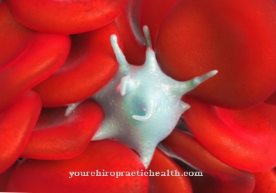
The primary hemostasis - hemostasis - takes place in 3 phases. The first step is platelet adhesion, followed by reversible platelet aggregation and the formation of an irreversible platelet plug.
The task of hemostasis is to repair injured vessels as quickly as possible so that blood loss remains as low as possible. Therefore, when the endothelium is injured, vasoconstriction occurs immediately. The constriction of the vessels also means that the blood flows more slowly.
This supports the next step: platelet adhesion. The blood platelets (thrombocytes) attach to subendothelial structures such as collagen. This accumulation is initiated directly by the collagen receptor and indirectly by the so-called von Willebrand factor. The adhesion activates the platelets and the reversible platelet aggregation is initiated. So the platelets accumulate close together, and ultimately an irreversible platelet plug is formed.
Function & task
The function of platelet adhesion is an interaction of the von Willebrand factor with various glycoproteins. At the molecular level, it is a ligand-receptor interaction. The ligand is the so-called von Willebrand factor and the most important thrombocytic receptor is the GP Ib / IX complex.
The accumulation of platelets on subendothelial surfaces is mediated by the GP Ia / IIa receptor complex - the collagen receptor. The von Willebrand factor (vWF) also has an indirect influence on this. This is a large glycoprotein that is released from the injured endothelium. It can form bridges between special membrane receptors of the platelets (GP Ib / IX complex) and collagen fibers. Fibronectin and thrombospondin are also involved in this bridge formation. The exposed collagen structures also interact without the vWF with GP Ia / IIa and GP VI on the platelet surface. Both reactions contribute to the fact that the platelets roll along the vessel wall and finally adhere.
In summary: The collagen receptor leads to a single layer of platelets. The von Willebrand factor causes the thrombocytes to adhere firmly via GP Ib / IX.
This platelet adhesion, in combination with the vasoconstriction, leads to an initial reduction in bleeding. It is also important for the activation of the platelets. The activation of the platelets also involves the release of adenosine diphosphate (ADP), fibrinogen, fibronectin, vWF and thromboxane A2.
The reversible platelet aggregation is initiated by the platelet activation. Platelets are tightly packed together via fibrinogen bridges. The vasoconstriction is additionally intensified by the escape of blood plasma into the interstitium. Thrombin causes the blood platelets to fuse into a homogeneous mass, the irreversible platelet plug. The formation of the irreversible platelet plug and vasoconstriction ensure that, in the event of minor injuries, temporary hemostasis occurs within a short time.
Primary hemostasis can be inhibited pharmacologically. For example acetylsalicylic acid (e.g. Aspirin®), which suppresses the synthesis of thromboxane A2. Additional platelet function inhibitors are ADP and GP IIb / III a antagonists. These drugs are often used temporarily when bedridden, such as before and after surgery, for example. They serve to inhibit blood clotting and thereby avoid thrombosis and embolism. This procedure is called thrombosis prophylaxis.
You can find your medication here
➔ Drugs for wound treatment and injuriesIllnesses & ailments
The adhesion tendency (adhesion) of the platelets can be measured using defined glass surfaces or on glass bead filters (retention). An inadequate function of the platelet adhesion manifests itself primarily in the increased tendency to bleed.
Platelet adhesion disorders are hereditary. They are based on a disturbed interaction between the platelets and the vascular endothelium. The cause of this disorder can, for example, be a deficiency of the von Willebrand factor, as is the case with Willebrand-Jurgens syndrome. This disease is inherited in almost all cases. Acquired forms have so far only been described very rarely. The severity and severity of the syndrome can vary. The disease often progresses very easily, so that the disease often goes unnoticed for a long time.
Roughly one can distinguish 3 types of the disease. Type I has a quantitative deficiency in the von Willebrand factor. This form is the most common, it shows very mild symptoms and often allows the patient to lead a normal life. Only the bleeding time is a little longer, and patients suffer more frequent secondary bleeding during operations. In type II, on the other hand, there is a qualitative defect in the Willebrand factor. This form is the second most common, but only affects 10-15% of all patients with Willebrand-Jürgens syndrome. Type III has a very severe course, but is the least common.
The disease is diagnosed in the laboratory if there are symptoms. The amount and activity of the von Willebrand factor is measured here. Long-term therapy is usually not necessary for diagnosis. Desmopressin, which increases the amount of the von Willebrand factor five-fold, is given to those affected only before operations.
The Bernard-Soulier syndrome, on the other hand, occurs much less frequently. The disruption of platelet adhesion is due to a hereditary defect in the membrane receptor for the von Willebrand factor (GP Ib / IX). This disease is also associated with an increased tendency to bleed. However, spontaneous bleeding is rare. The diagnosis is again made in the laboratory and therapy is rarely necessary due to the mild symptoms. Patients only have to be careful not to take any platelet aggregation inhibitors such as Aspirin®. These can lead to serious bleeding complications. Platelet concentrates are only substituted in acute cases, such as after major blood loss.







.jpg)

