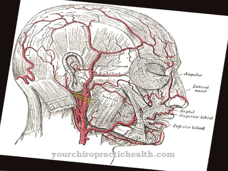
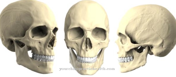
The paired Maxillary artery represents the natural continuation of the external carotid artery from the departure of the superficial temporal artery. The maxillary artery can be divided into three sections and in its end area forms connections to other arterial vessels that arise from the facial artery. Their task is to supply some of the organs and tissues located in the deep facial region.
What is the maxillary artery?
The maxillary artery, also called the Maxillary artery denotes, represents the natural continuation of the external carotid artery or external cervical artery. The external carotid artery divides into the two branches, arteria temporalis superficialis (superficial temporal artery) and arteria maxillaris (maxillary artery).
It is a paired artery that is mirror-inverted on both sides of the head. From the artery, which can be divided into three sections, numerous smaller arteries branch off to supply their target organs or tissue. Target organs and target tissue are, for example, the lower jaw, teeth and tympanic cavity of the middle ear as well as the dura mater of the brain and spinal canal. In its end branches, the maxillary artery forms so-called anastomoses, connections to side branches of the facial artery (facial artery).
Anatomy & structure
The maxillary artery embodies the transition form from the elastic to the muscular type of an artery. This means that it perceives the passive properties of the large, elastic arteries close to the heart to a certain extent, but also has the active mechanism of changing the lumen by tensing or relaxing the smooth muscle cells in its walls.
The lumen change is mainly controlled hormonally via sympathetic stress hormones (tension) and parasympathetic inhibitors of the stress hormones (relaxation). The maxillary artery is one of two terminal branches of the external carotid artery (external carotid artery) and arises in the retromandibular fossa at the level of the transition from the neck to the head. The maxillary artery is divided into three sections, the mandibular, pterygoid, and pterygopalatine parts.
A total of five arteries arise from the mandibular section, which run into the deep ear regions, into the tympanic cavity and to the lower teeth and run to certain areas of the hard meninges (dura mater). From the pars pterygoidea, also known as the intermuscular section, arise four arteries that mainly supply the masticatory muscles and the cheeks. Five arteries branch off from the pars pterygopalatina and supply the palate, nasal cavity and teeth of the upper jaw.
Function & tasks
The maxillary artery is part of the arterial side of the great blood circulation and thus contributes, in conjunction with the rest of the arterial network, to smoothing the blood flow and maintaining diastolic blood pressure. The elastic walls stretch a little during the systolic blood pressure peak and contract again during the diastole, the relaxation phase of the heart chambers, so that they make a small contribution to the passive Windkessel effect of the large arteries close to the heart.
Due to the musculature in the arterial wall, which surrounds the artery partly in a ring and partly in a spiral, the maxillary artery also makes a contribution to adapting and controlling the blood pressure to different performance requirements. In its ostensible main function, the maxillary artery serves to supply certain facial regions and deeper tissues with fresh, oxygen-rich blood. Specifically, the side branches of the maxillary artery carry oxygen-rich blood to the upper and lower jaw, the masticatory muscles, the nasal cavity and the tympanic cavity of the middle ear. In addition, parts of the dura mater, the hard meninges, and the palate are supplied by branches of the maxillary artery.
The fact that some terminal branches of the maxillary artery are connected to other arteries, i.e. form so-called anastomoses, shows that the maxillary artery with its branches is of enormous importance. If pathological occlusions occur, the connected arterial network can serve as a back-up and prevent necrosis of the affected tissue.
If there are direct connections between the arterial and venous parts of the blood circulation without the interposition of the capillary system, the problem is usually pathological arteriovenous malformations that can lead to serious clinical pictures. In certain cases, such a short circuit between the arterial and venous vein system can also be brought about artificially for the treatment of certain diseases.
You can find your medication here
➔ Medicines for visual disturbances and eye problemsDiseases
The maxillary artery is subject to the same conditions that apply to the other arteries with regard to its potential risk from diseases. No specific disease of the maxillary artery is known.
The most common problems arise from disorders of the blood flow, which can be triggered by constrictions, stenoses, in the lumen of the maxillary artery. The most common reason for a stenosis is arteriosclerosis, a penetration of the arterial wall with plaques, deposits that make the arterial walls inelastic and cause constrictions in the artery or block it completely. Inflammatory reactions can occur where plaques are deposited in the arterial wall. The inflammatory reactions can trigger the formation of blood clots and lead to a complete occlusion of the artery, a thrombosis.
This can have far-reaching consequences because the affected tissue areas can no longer be supplied with oxygen-rich blood. In rare cases, infectious and inflammatory damage to the vascular wall can cause a bulging, aneurysm, to form in the maxillary artery, which creates the risk of internal bleeding. If an aneurysm forms in the area of the dura mater, there is a risk that the bulge will lead to compression processes in the brain and to impairment of certain brain functions. In very rare cases, the maxillary artery can be affected by an embolism. The embolism is triggered by a thrombus, which is accidentally washed into an artery by the bloodstream and leads to an occlusion of the vessel if its diameter is less than that of the thrombus.

