Burkholderia pseudomallei is a bacterium from the Proteobacteria division and from the Burkoholderiaceae family. It can cause melioidosis in humans.
What is Burkholderia pseudomallei?
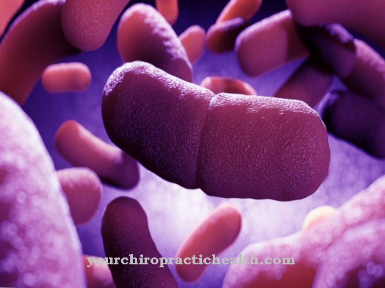
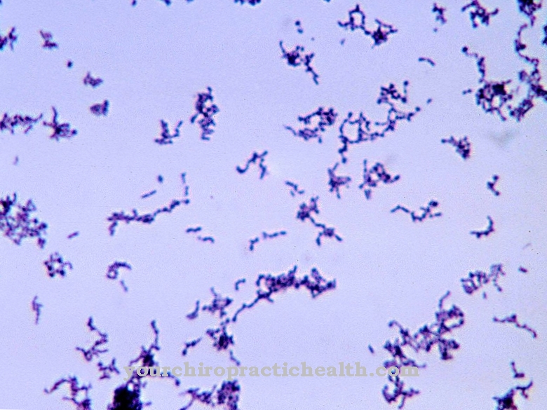
The pathogen Burkholderia pseudomallei is one of the gram-negative bacteria. Gram-negative bacteria can be stained red in the so-called Gram stain. In addition to a thin layer of peptidoglycan made from murein, the gram-negative bacteria also have a cell membrane on their outer shell.
Burkholderia pseudomallei is strictly aerobic. Aerobic bacteria need oxygen for their metabolism. The bacterium is rod-shaped and thus belongs to the rod bacteria. It lives saprophytically. Saprophytes are organisms that feed on dead organic matter. They break down these energy-containing substances and then convert them into inorganic substances. In the case of bacteria in particular, the transition from a saprophyte to a parasite is fluid.
Burkholderia pseudomallei grows intracellularly and is oxidase positive. The microbiological process of the oxidase reaction tests whether the corresponding bacterial strain has the enzyme cytochrome C oxidase. This information plays a decisive role, among other things, when choosing a therapy.
Burkholderia pseudomallei comes from the genus Burkholderia. However, this classification did not take place until the 1990s. The bacterium was previously assigned to the groups Bacillus, Mycobacterium, Peifferella, Actinobacillus and Pseudomonas.
Burkholderia pseudomallei has an average diameter of 0.6 μm and becomes approximately 5 μm long. It moves with the help of flagella. Flagella are also known as flagella. These are thread-like structures that sit on the surface of the bacteria and are used for locomotion.
Occurrence, Distribution & Properties
Burkholderia pseudomallei is found in the ground and in water. Pets and wild animals also serve as reservoirs. The bacterium is endemic to both Northern Australia and Southeast Asia. Serotypes are also differentiated on the basis of the geographical areas. The / ara + serotype is more likely to be found in Southeast Asia. Serotype II / ara is preferred in Northern Australia.
Burkholderia pseudomallei is mainly infected through direct contact with contaminated soil or water.In tropical countries workers in rice fields are often infected with melioidosis. The pathogen enters the organism through the smallest skin damage. The infection can also take place via inhalation or oral intake. A person-to-person infection is also possible through body fluids. Furthermore, there is a risk of infection in the laboratory through the inhalation of infectious aerosols.
There are always cases in the news where the bacterium escaped from laboratories. The last time this happened was in 2014 in the US state of Louisiana. There, four rhesus monkeys fell ill in an outdoor area and a scientist was also infected. Burkholderia pseudomallei is considered a potential bio-weapon and is on the bio-weapons agent list.
Illnesses & ailments
The bacterium Burkholderia pseudomallei causes the infectious disease melioidosis. This is also known as Whitmore's Disease or Pseudorotz. The incubation period is very different. It can last as little as two days or several years. The course and symptoms of the disease are also very different.
Many infections are completely asymptomatic. Mild chronic illness develops in other patients. Still other sufferers react with an acutely fulminant disease. After the pathogen enters the body through a skin lesion, a small nodule often develops in the skin. The surrounding lymph vessels become inflamed (lymphangitis) and the lymph nodes also react (lymph node swelling). Patients have a fever and feel tired, limp and sick.
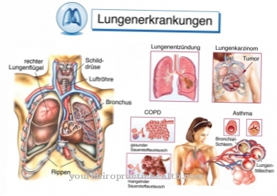
This local infection can quickly spread to the whole body. In this case it is a generalized, septicemic form. In this life-threatening course, abscesses form all over the body. The lungs are also affected by abscess formation. The patients suffer from impaired consciousness and severe shortness of breath. The breathing rate is increased. If the pathogen did not get into the body through the skin, but was inhaled, pneumonia usually develops directly.
A distinctive cavern formation is characteristic of melioidosis. Caverns are pathological cavities within the lungs. Gas exchange can no longer take place in these cavities, so that the functionality of the lungs is severely restricted. Often a pleural effusion develops in addition to pneumonia. This causes fluid, in most cases inflammatory exudate, to enter the pleural space. The compression of the lungs makes breathing even more difficult.
In many cases, melioidosis is chronic and has no fever. Abscesses form in various organs. Depending on the affected organ system, different symptoms can occur. Diabetics and people with a suppressed (suppressed) immune system are particularly at risk. Even if there are no symptoms for several years after an infection, the disease can manifest itself in the case of an immune deficiency.
Antibiotics and chemotherapy drugs are used in high doses to treat melioidosis. These are usually given intravenously. After the acute symptoms have subsided, the therapy often has to be continued orally for several months. Abscesses caused by the disease are surgically removed.
There is no effective prophylaxis against the Burkholderia pseudomallei bacterium. Anyone traveling in endemic areas should carefully clean and disinfect skin injuries. Burkholderia pseudomallei is sensitive to various disinfectants.







.jpg)


.jpg)



.jpg)
