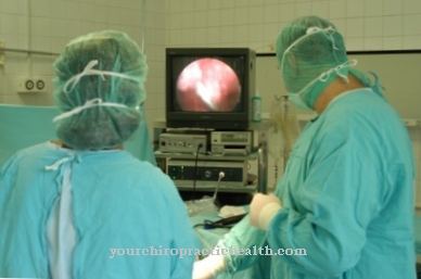
With the help of Chorionic villus sampling the unborn child can be examined for possible genetic disorders during pregnancy. This examination method can be carried out at a very early stage in pregnancy.
What is the chorionic villus sampling?

The prenatal diagnosis with the help of chorionic villi was first described in 1983. It is an invasive examination method with which chromosomal abnormalities can be detected and certain metabolic diseases can be identified.
A chorionic villus sampling is advisable if there are any abnormalities on the ultrasound, the presence of chromosomal abnormalities in the parents or if certain inherited diseases are suspected. However, it is not a routine examination. The chorionic villus sampling is only carried out after appropriate information and with the consent of the pregnant woman.
If the unborn child is suspected or at risk of illness, the health insurance company bears the costs of the examination. At the request of the parents, the chorionic villus sampling can also be performed in other cases at your own expense. However, a risk-benefit assessment should be made prior to the procedure. An increased likelihood of trisomy 21 in the child is present, for example, in pregnant women aged 35 and over and justifies the diagnosis by means of chorionic villus sampling.
Function, effect and goals of the chorionic villus sampling
The chorionic villus sampling can be used as a diagnostic method in the first trimester of pregnancy. From the eighth week of pregnancy, an analysis of the cells from the so-called chorion is possible. The method enables the child's chromosomes to be examined at the earliest possible point in time during pregnancy when other examinations such as amniocentesis are not yet possible.
In the past, doctors used chorionic villus sampling as early as the ninth week of pregnancy. However, the chorionic villi biopsy should not be performed before the 11th week of pregnancy and is usually not performed earlier today. The chorion is a layer of cells on the outside of the amniotic sac. This cell layer is evenly covered with villi, protuberances on the surface.Even if this is not the cell material of the unborn child, it is genetically identical to the unborn child and is therefore suitable for diagnostics.
The chorionic villi form part of the placenta that supplies the unborn child with nutrients and oxygen. In a chorionic villus sampling, chorionic villi are removed from the womb. This is done either with the help of a hollow needle, which the doctor inserts under ultrasound control through the abdominal wall into the placenta, and there cell material is removed by puncture. Another possibility is the removal of chorionic villi via a catheter that passes through the vagina and through the cervix into the placenta.
Collection via the cervix is seldom done because of the higher risks. In some cases, however, a chorionic villus sampling is not possible for anatomical reasons. For the subsequent genetic examination in the laboratory, 20 to 30 milligrams of chorionic villi are required. A chromosome image, the so-called karyogram, is created from the cells removed. In special cases, a DNA analysis can also be carried out after prior genetic advice. This allows the unborn child to be examined for molecular genetic diseases, such as various muscle diseases. With the help of the karyogram, various genetic characteristics can be detected.
These include trisomy 21, which is known as Down syndrome, trisomy 13 or Patau syndrome, trisomy 18 or Edwards syndrome and trisomy 8. Some metabolic diseases can also be analyzed using the karyogram. The first results of the laboratory test are available after a few days. If the findings are unclear, a long-term culture of the removed cells is required, the results of which are available after approximately two weeks.
The chorionic villus sampling is only performed at specialized centers and can therefore usually not be performed by a resident gynecologist. The aim of the investigation is to detect or rule out genetic diseases as early as possible in pregnancy in order to avoid medical risks and psychological stress from a late termination of pregnancy. In advance of a chorionic villus sampling, parents should be informed about the lack of therapeutic options for curing most of the diseases that can be identified by this examination. If a genetic disease is detected, the only remaining option is usually to accept the child with the disease or to terminate the pregnancy.
Risks, side effects & dangers
A chorionic villus sampling increases the risk of a miscarriage. This examination method is therefore mainly chosen when the risk of miscarriage is lower than the probability of a chromosome abnormality or the presence of a hereditary disease.
There is also a low risk of deforming the child's extremities from the procedure. Vascular injuries or bleeding as well as infections can also occur in rare cases after a chorionic villus sampling. In rare cases, which are around two percent, a misdiagnosis can occur. The chorionic villi can differ genetically from the child's cells. Likewise, cells within the placenta can in rare cases differ genetically from one another. This is known as the placenta mosaic.
As a result, examined cells can show a trisomy, even though the unborn child has a normal set of chromosomes. A trisomy can also go undetected during the examination. If the results are positive, it is sometimes recommended to confirm the test result by performing a further test, such as an amniotic fluid test. In the meantime, the result of a fine ultrasound in the 13th week of pregnancy is often waited for before a chorionic villus sampling. Depending on the findings and the assessment of the risk for a trisomy, a decision can then be made to perform a chorionic villus sampling.












.jpg)

.jpg)
