With the Defibrillation First aiders use a direct current impulse to remedy a life-threatening cardiac arrhythmia which, if not counteracted in good time, can result in a fatal heart attack. Defibrillation takes place exclusively through a successful shock application. The synonym for defibrillation is Defibrillation.
What is defibrillation?

The direct current pulse on the patient is given by shock. The defibrillator acts as a shock generator for defibrillation and cardioversion. It is a controlled delivery of electric shocks to the heart muscle. The European Resuscitation Council (ERC) defines the absence of the original cardiac arrhythmia five seconds after the shock is given as successful defibrillation.
Defibrillation is performed in the event of resuscitation in the case of cardiac arrhythmias such as ventricular fibrillation, ventricular flutter and pulseless ventricular tachycardia (life-threatening rhythm disturbance emanating from the ventricles). In the meantime, so-called AED defibrillators are increasingly being used. These devices take over the ECG diagnosis and guide through the measures for cardiopulmonary resuscitation using optical and acoustic signals.
Function, effect & goals
The contraction, the contraction of the heart muscle, occurs through the depolarization (discharge) of the muscle fibers, whereby the repolarization is an electrical phenomenon in which the heart's original state of charge is restored. Cardiac arrhythmias and thus sometimes life-threatening conditions that can lead to fatal heart attacks always occur when the heart muscle cells no longer work in a coordinated manner and the blood supply to the body is not guaranteed.
The heart remains active, but does not show an orderly pumping function. Clinically, the first signs of a life-threatening cardiac arrest are showing. If the patient is in such a situation, the doctors use an EKG to check the underlying heart rhythm. Based on this data, the cardiologists decide whether a shockable rhythm is present or not. To treat a patient with life-saving defibrillation, first responders place one electrode over the top of the heart and a second over the base of the heart.
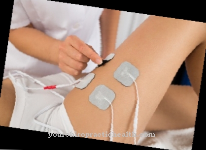
The electrodes are set using adhesive electrodes or so-called paddles. Paddles are large-area plate electrodes, which, in contrast to adhesive electrodes, require less time to attach. The paddles are attached to the right, parasternal below the clavicle (collarbone) and to the left at the level of the fifth intercostal space (space between two adjacent ribs) in the anterior axillary line. In the case of ventricular tachycardia (ventricular fibrillation), the position of the paddles is swapped in the so-called cross-check in order to rule out any disturbances in the ECG that can simulate a shockable rhythm, even though there is, for example, an asystole (lack of contraction of the heart muscle).
An ideal situation is when the cardiac rhythm massage is only interrupted for a very short period of time, less than five seconds, before the shock is given. In the case of so-called manual defibrillators, however, this is only possible with a well-rehearsed and experienced team. Then the doctors try to depolarize as large a mass of the heart muscle cells as possible, they are set to "zero". This life-saving measure completely interrupts the states of excitement that were previously circulating in the ventricle (one of the two lower chambers of the heart) and the heart now has the chance to allow the excitation to take place again in a natural process (conduction system).
If the defibrillation is successful, the sinus node (primary pacemaker center of the heart) takes control of the work of the heart muscle again. However, shock alone is not sufficient. The medical professionals must then continue with manual resuscitation in order not to "lose" the patient. There is no time to feel the pulse or look at the ECG monitor, all measures must be taken very quickly.The myocardium (heart muscle that makes up most of the heart wall) needs some time to recover from the stress that this life-threatening situation brings with it.
The electrical cardioversion is not a regular emergency measure and is usually ECG-controlled, whereby the direct current surge into the non-vulnerable phase (period in which an extraordinary impulse does not trigger ventricular fibrillation or ventricular flutter during the cardiac cycle) of the heart's action is triggered. It is used for atrial fibrillation and (supra) ventricular tachycardia. The ideal situation is when a resting ECG is recorded in addition to the ECG lead II, which is performed using the device paddles on the sternum (breastbone) and appex (apex of the heart).
Cardioversion is performed using R-wave synchronous electric shocks, a significant difference to defibrillation that is not performed synchronously. The synchronous delivery of the electric shocks means that the current delivery is triggered by the user, but the device delays it until the R-wave can be closed again. With this method, the medical professionals avoid that the current output during the refractory phase (relaxation phase) follows the spread of excitation.
If a current were to be delivered during this phase, there is a risk of ventricular fibrillation and cardiovascular arrest. The electrical cardioversion works with a lower Joule strength (50-100) than the defibrillation. Cardioversion requires patients to be given a benzodiazepine (midazolam) and a hypnotic (etomidate).
You can find your medication here
➔ Medicines for cardiac arrhythmiasRisks, side effects & dangers
In the case of contraindications and unfavorable environmental conditions, defibrillation can be dangerous. A contraindication is present if the patient has a body temperature of less than 27 degrees Celsius, i.e. severe hypothermia. Further contraindications are digitalis intoxication (poisoning by digitalis), existing thrombi with the risk of embolism, hyperthyroidism (pathological overactive thyroid) and changed heart morphology.
The environmental conditions are unfavorable and therefore risky when the surface is wet or there is metallic contact between the patient and the first aider. Defibrillation must also be avoided in the event of a risk of explosion. If the patient has issued an advance directive against any resuscitation measures, the medical professionals must refrain from defibrillation. During both defibrillation and cardioversion, nobody is allowed to touch the patient or the bed, as the electric shock can be transmitted to these people and put their lives at risk. Due to the risk of burns, the patient must not wear any metallic objects such as rings or belts.
Dental prostheses are also dangerous, as they can interrupt the spasm triggered during resuscitation or obstruct breathing if loosened. Due to the risk of aspiration, the patient must be fasted during cardioversion. With electrical cardioversion, the patient is anticoagulated three weeks before and three weeks after the treatment (a drug is given to prevent blood clotting). Possible complications can include pulmonary embolism due to the detachment of thrombi, additional cardiac arrhythmias, anaphylaxis (allergic reaction to the administration of medication) and skin reactions in the area of the electrodes.





.jpg)

.jpg)




.jpg)
