The term Ectodermwhich is derived from the Greek ektos outside and derma, the skin, denotes the first upper cotyledon. In the course of development it forms the nervous system as well as the skin in humans and also in the animal world.
What is the ectoderm?
During the so-called gastrulation, which is an essential part of development, the blastula, which consists of a single layer of cells, becomes a structure consisting of three different layers of cells.
The blastula is the egg cell after fertilization by a sperm and after multiple cell division. These three cell layers that make up the blastula after gastrulation are called the ectoderm, the outer cell layer, the mesoderm, the inner cell layer, and the endoderm, the inner cell layer. Later in development, the ectoderm forms the nervous system, sensory organs, skin and teeth.
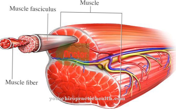
The mesoderm develops into the muscle tissue, the skeleton, the blood vessels and the connective tissue. The endoderm, on the other hand, forms the epithelium, liver, pancreas, as well as the respiratory and digestive systems after the embryo has developed completely. These three cell layers are also known as the cotyledons and are the basis from which the organs of humans and animals develop.
Anatomy & structure
The cotyledons each consist of a layer of cells. However, the cells of the cotyledons, including the ectoderm, are not yet specialized. They are preprogrammed to develop into a certain type of cell. This is described as differentiation.
This differentiation is controlled. Each cell contains the information in which cell type it should develop. The cells of the different cotyledons have different information for differentiation. Even within a cotyledon, the cells have different information for differentiation. Therefore, different cell types are formed from each cotyledon.
Like the ectoderm which forms the nervous system, but also the teeth. The cells of the cotyledons are thus determined, they have a predefined path of differentiation. It is possible, however, for cells from one cotyledon to become cells of another cotyledon. This happens when the mesoderm is formed. This is then called transdetermination of the cell. It changes its original determination.
Function & tasks
Animals and thus also humans, which form the three cotyledons, are called bilaterally symmetrical animals. The blastula, or in human sore higher mammals, it is also called the blastocyst, is a kind of hollow sphere, which consists of a layer of cells. It initially develops into a gastrula.
The two primary cotyledons are formed. These are the external ectoderm and the internal endoderm. In this stage of development, the endoderm forms the original mouth and the so-called original intestine. The mesoderm is formed a little later. During gastrulation, the cells are rearranged. The cavity inside the ball is filled more and more while the ectoderm closes the entire outside of the gastrula. The gastrulation then changes into the neurulation. This is the formation of the neural tube. The neural tube later forms the central nervous system when the development process is complete.
The neural tube is formed by reshaping the neuroectoderm. This forms from the ectoderm and then forms the neural tube by folding over the cell layer. First, the ectoderm thickens, which is induced by specific signals from the mesoderm. The neural plate forms. The edges of these plates form the neural bulges and form the neural groove between them. These neural bulges and the neural groove then form the neural fold, which finally closes to form the neural tube. The front area of the neural tube forms you towards the brain and the tube behind it then forms the spinal cord.
The cavity of the neural tube fills with cerebrospinal fluid. In addition, the eye vesicles are also formed in the front area, which later become the actual eyes. This process is known as the primary neurulation. Secondary neurulation, on the other hand, is the formation of fluid-filled cavities in the areas that adjoin the neural tube.
You can find your medication here
➔ Medicines against redness and eczemaDiseases
Spina bifida is the malformation of the neural tube. This malformation can take different forms. It occurs between the 22nd and 28th day of embryo development. During this time, neurulation takes place, i.e. the formation of the neural tube by the neuroectoderm.
Spina bifida refers to the incorrect closure or failure of the neural tube in the rear part of the neural tube. The spina bifida shows itself in different forms. The spina bifida occulta is characterized by an absence of the membranes of the spinal cord, the meninges. This form of spina bifida is not externally recognizable.This form is not severe and does not require treatment. The spina bifida aperta, on the other hand, is characterized by a neural tube that is not completely closed. There are three forms of spina bifida aperta. Meningocele is a mild form of this disease.
The membranes of the spinal cord bulge out and form cysts under the skin, which can be surgically removed without affecting the spinal cord. Meningomyelocele is a severe form of spina bifida. The spine has one or more fractures through which parts of the spinal cord protrude from the spine. The nerves are damaged. However, this can be treated surgically. Myeloschisis refers to the case that nerve tissue is completely exposed. This is the most severe case of spina bifida aperta.





.jpg)


.jpg)



.jpg)
