As embryonic head development the development of the skull, the differentiation of the pharyngeal arch systems and the development of the craniofacial system are summarized. The development of the skull primarily forms the bony base of the skull, while the pharyngeal arches form organs. Developmental disorders cause dysplasias (visible malformations).
What is embryonic head development

The head development of the embryo is a multi-phase process, during which the head and its structures also develop the embryonic neck. The development phases correspond to the development of the skull, the differentiation of pharyngeal arches and the differentiation of the craniofacial system. The basic elements for the embryonic attachment of the head and neck are the pharyngeal arches and the para- and prechordal cartilages that are attached to the uppermost somites.
The development steps are carried out on a genetic basis. The responsible genes are linked to the Homeobox genes. For the skull itself, the neural crest, the paraxial mesoderm, the occipital somites and the two upper pharyngeal arches are relevant as starting materials. The chewing apparatus, the ossicles, the facial muscles, the hyoid bone, the larynx and parts of the arteries are differentiated from the pharyngeal arches. The development of the craniofacial system corresponds to a facial structural development from the previously created facial bulges.
Function & task
There is a close connection between the development of the skull capsule and the development of the meninges. In the sixth week of embryonic development, the system for the brain is surrounded by compacted mesenchymal cells. The outer leaf is compressed into the dura mater encephali. The leptomeninx arises from the inner leaf. In the section of the brain base, the meninx primitiva become the pre-cartilage cells of the chondrocranium. In addition, desmocranium osteoblasts are formed. The preformed part of the skull is cartilaginous and is called the chondrocranium. After ossification, this section corresponds to the base of the skull.
Part of the skull is mesenchymal. This so-called desmocranium is ossified to form the roof of the skull and forms a large part of the bones that lie in the viscerocranium. The squama occipitalis and the pars squamosa ossis temporalis have chondral and desmal origins.
The base of the skull is created in the course of embryonic development, primarily through processes of chondral ossification that take place on the chondrocranium. The skull has its origin in desmal ossification based on the desmocranium. The cartilaginous base of the skull is formed from material from the notochord. The basis of this is formed by prechordally paired central cartilages and their lateral cartilage pairs of alae temporales and orbitales.
The basal plate of the skull arises at the anterior end of the notochord. The pair of ear capsules, which later receive the inner ear, are created on the opposite side. The basal plate is connected to occipital somites, which are involved in the development of the foramen magnum. Remnants of cartilage from the ossification centers remain in the clivus until puberty. Some parts of the skull remain cartilaginous for life, such as the nasal septum.
In the area of the desmocranium, an opposing interaction of bone-forming osteoblasts and bone-degrading osteoclasts builds up, which enables widespread ossification. This is how the complicated shape and length relationships of the individual skull bones can arise.
Sutures are the contact points of the growing skull plates that create bone sutures. The sutures usually ossify postnatally. The roof of the skull can therefore expand according to its shape. Large cover bone plates and connective tissue gaps, known as fontanelles, can be seen at the contact points in newborns.
The pharyngeal arch differentiation follows these cranial processes. Development begins at four or five weeks of age. In the fifth week there are four ectodermal depressions in the ventrolateral head area, which are called gill furrows. Four pharyngeal pouches of the endoderm grow on the inside towards these gill furrows. These processes divide the mesodermal tissue into four pharyngeal arches. The caudal, fifth pharyngeal arch is poorly differentiated and soon recedes. All pharyngeal arches become cartilage elements or muscle structures, each of which is assigned a pharyngeal arch nerve and a pharyngeal arch artery.
The endodermal inner pharynx forms individual organs of the head and neck region. Among the ectodermal outer throat furrows, only the first one becomes an organ that becomes the external auditory canal and part of the eardrum. The neck cavity migrates in a caudal direction and migrates in the direction of the second pharyngeal arch, so that a cavity with reclosing forms on the lateral neck.
The subsequent development of the craniofacial system focuses on the application of the facial bulges. The forebrain vesicles expand and, together with the first pharyngeal arch and the heart bulge, delimit the head area and the mouth of the child. The oral cavity is closed by the oropharynx membrane, which later tears and connects the foregut with the amniotic cavity. In the fourth week, an ectodermally covered cushion of mesenchyme forms, from which the medio-cranial forehead nasal bulges and the upper and lower jaw bulges extend.
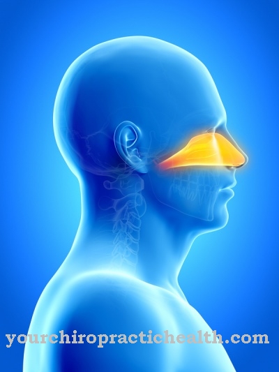
The first differentiation of the facial bulges occurs through an ectodermal thickening, which gives rise to the olfactory placode at the ends of the forehead and nose bulges. The proliferation of the mesoderm turns it into the olfactory pits and the olfactory sacs and also divides a middle from a lateral nasal bulge on both sides. Then the tear-nasal furrow separates the lateral nasal bulge from the upper jaw bulge. The injection of the surface epithelium supports the development of the lacrimal sac and nasal duct. The nostrils are formed from the lateral nasal bulges.
The intermaxillary segment is formed by the middle nasal bulges growing towards each other and fits into the paired upper jaw palatal anlage. After the elements grow together, a bridge of the nose forms. The incisive canal remains open as a suture.
The eye systems experience a frontalization. The appendages for the outer ear migrate from the neck area in the cranial direction. At the same time, the maxillary bulge pushes past the lateral nasal bulge and merges with the middle nasal bulge. The lateral upper lip, the upper jaw and the paired secondary palatal anlage arise from the upper jaw bulge. The medially fused maxillary bulges create the base of the lower lip and the desmal mandible. The lateral jaw and upper ridges fuse so that the wide stomatodeum opening narrows into a defined mouth.
Illnesses & ailments
Embryonic development disorders from the fourth week of embryonic development can cause various malformation syndromes by disrupting head development. Some of these disorders are genetic and mutation related. Others are favored by external factors such as exposure to toxins or malnutrition during pregnancy.
Disturbances in the desmocranium development can, for example, correspond to an early ossification of the individual sutures. This phenomenon is known as craniosynostosis and gives rise to deformed skull shapes, such as the tower skull, the pointed skull, the punt skull, the triangular skull or the crooked skull. Some cranial dysplasias are associated with mental developmental delays or mental retardation, such as the premature ossification of all sutures, which constricts the patient's brain and prevents it from expanding.
If the developmental disorder does not correspond to a disorder of the skull development but to a disorder of the pharyngeal arch development, serious symptoms can also arise. Remnants of the lateral cervical sinus can develop, for example, neck fistulas that reach down into the pharynx or end blindly.
Other symptoms are present in actual malformation syndromes such as Goldenhar syndrome, which causes oculo-auriculo-vertebral dysplasia. This syndrome is caused by combined anomalies in the first and second pharyngeal arches and is associated with conduction symptoms with an underdeveloped jaw and hypoplastic ear region.
These malformations are associated with dysplasias of the cervical spine. Disturbed development of the craniofacial system can also result in obvious malformations. For example, if the middle nasal bulges only incompletely fuse with the maxillary bulge, a cleft lip and palate is created. The cleft formation disorders can result in abnormalities such as the transverse facial cleft or the lower lip cleft. The clinical picture of disorders during embryonic head development is accordingly varied.











.jpg)
