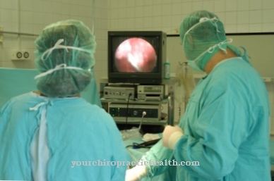
The endoscopic retrograde cholangiopancreatography (ERCP) is an imaging procedure based on X-rays. It is used to visualize the biliary and pancreatic ducts. This method is an invasive diagnostic procedure and therefore also involves risks.
What is endoscopic retrograde cholangiopancreatography?

Endoscopic retrograde cholangiopancreatography is often performed if the biliary tract or pancreatic disease is suspected. This is an invasive diagnostic procedure that works with the help of X-rays.
With this procedure, pathological changes in the area of the biliary and pancreatic ducts can be detected. It is only used if the examination using magnetic resonance cholangiopancreatography (MRCP) does not provide any clear diagnostic results. In contrast to ERCP, MRCP is a non-invasive procedure. Sometimes this method does not detect all changes.
However, if there are undiagnosed changes in this area, these can be clearly shown by the ERCP. In addition to the diagnostic examinations, minor surgical interventions are also performed if necessary. The term "endoscopic retrograde cholangiopancreatography" denotes the use of an endoscope which, retrograde, i.e. from the exit, introduces a probe into the bile or pancreatic ducts using contrast media and depicts this area there.
Function, effect & goals
Endoscopic retrograde cholangiopancreatography is used for suspected gallstones, narrowing of the bile ducts due to inflammatory changes or tumors of the bile duct and for chronic inflammation, cysts or tumors of the pancreas. It is an invasive examination method that uses X-rays to depict the biliary and pancreatic ducts.
Due to the existing risks from radiation, contrast media and invasive surgery, this method is only carried out if MRCP and ultrasound examinations have not produced any results. Small surgeries can also be performed during ERCP if necessary. This concerns the taking of tissue samples, the expansion of the mouth of the duct systems, the expansion or bridging of constrictions with stents. The procedure of an endoscopic retrograde cholangiopancreatography is similar to a gastroscopy. An endoscope attached to a tube is inserted through the mouth and into the duodenum beyond the stomach.
There, contrast medium is injected into the father's papilla against the outflow direction of the bile and the pancreatic secretions (retrograde) and a probe is extended from the endoscope. The probe is then introduced into the biliary or pancreatic ducts via the Father's papilla. The Father's papilla represents the common exit of the biliary and pancreatic ducts. At the end of the device there is a light source and a camera. This can be used to make this area visible. The probe (catheter) uses X-rays to record the interior of the bile duct and pancreatic duct and can thus detect stones, constrictions or tumors.
Small interventions can also be carried out if necessary. It can happen that the father's papilla is too narrow and thus causes a bile drainage blockage. The papilla opening can be widened using the endoscope. To do this, it is cut open using a special catheter with an electrically moved wire. If the ducts are narrowed due to inflammation or tumors, so-called stents made of plastic or metal tubes are often placed in order to ensure the drainage of bile and pancreatic secretions again. The bile duct can also be examined with a sonographic probe. This method is called intraductal ultrasound. Gallstones that are close to the bile outlet can also be removed with the endoscope.
The main concern of ERCP is to diagnose gallstones, biliary tract cancer, inflammation of the biliary tract, pancreatic cancer, and unexplained bile drainage disorders. The advantage of endoscopic retrograde cholangiopancreatography is the detection of changes in the biliary and pancreatic ducts without the need for open surgery. A purely diagnostic ERCP can therefore also be carried out on an outpatient basis.
Risks, side effects & dangers
Endoscopic retrograde cholangiopancreatography detects undetected changes in the area of the biliary and pancreatic ducts very well. However, like any invasive procedure, it also involves certain risks. The examination is carried out under a short anesthetic. As with any anesthesia, the usual anesthetic risks can arise.
In advance, it must be clarified with the patient whether there may be allergies to certain anesthetics and contrast media. The contrast agent may irritate the bile ducts and the pancreas. Therefore, in rare cases, pancreatitis can develop. Injuries to the larynx, esophagus, etc. Gastrointestinal wall with corresponding bleeding occur. The risks of X-rays must also be considered. Therefore, this method should only be used when there is no other way to make a meaningful diagnosis. This procedure is particularly not recommended for pregnant women because the unborn child is at risk from the effects of X-rays.
In advance of the procedure, it is important that the patient is informed about the risks. In this conversation, important questions about allergies, previous illnesses or the use of medication should be clarified. Drugs that thin the blood can increase the risk of bleeding during this procedure. Therefore, it must be clarified with the doctor in which framework the examination can still be carried out. The risk of bleeding may not be as high, or it may be possible to temporarily stop taking blood thinners. For the examination to be successful, it is also important that there are no food residues in the digestive tract. Therefore, before the ERCP, patients should urgently comply with the doctor's instructions on food abstinence of at least six hours.







.jpg)

.jpg)
