Of the Flexor reflex is a self-reflex of the finger flexors, which is triggered by a blow on the palmar side of the distal phalanx of the middle finger. Excessive reflex diffraction is considered an uncertain pyramidal orbit sign or a sign of vegetative dystonia. The final clarification takes place via imaging and CSF diagnostics.
What is the finger flexion reflex?

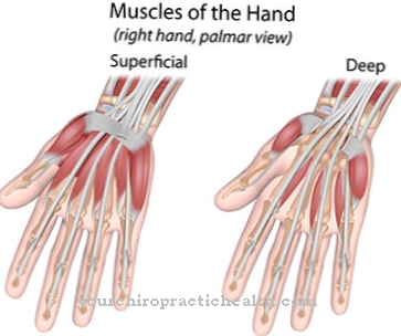
The hand has various flexor muscles. Two of these flexors are the flexor digitorium profundus muscle and the flexor digitorium superficialis muscle. These are the deep and superficial flexors of the fingers. The muscle reflex of these finger flexors is called the finger flexor reflex.
The reflex flexion movement is triggered by a blow on the palmar side of the distal phalanx of the middle finger and corresponds to finger flexion. The monosynaptic self-reflex was discovered in the 20th century by the German neurologist Ernest L. O. Trömner.
The nerves involved are the spinal cord segments C7 and C8 as well as the median and ulnar nerves. The finger flexion reflex is in itself a physiological self-reflex close to the threshold. In the case of an exaggerated or only one-sided reflex, on the other hand, we speak of a Trömner sign with pathological value. Unlike the Trömner reflex, the Trömner sign can be assessed as an indication of lesions in the pyramidal trajectories (albeit as uncertain) and thus corresponds to a weak pyramidal trajectory sign.
The pyramidal tracts are the spinal cord tracts between the upper and lower central motor neuron and are an important switching point for all voluntary and reflex motor skills.
Function & task
Muscle reflexes are monosynaptically interconnected protective reflexes that protect the skeletal muscles from overstretching. They are triggered by blows to the tendons in which the muscle spindles of the respective muscles sit.
Muscle spindles are extroceptive receptors. They detect strain and convert these mechanical stimuli into bioelectrical information. The non-contractile middle of a muscle spindle fiber is wound with afferent sensitive nerve fibers. These fibers are known as Ia fibers and convey excitation to the central nervous system.
When a muscle is stretched, the muscle spindle is stretched at the same time. The Ia fibers conduct this stimulus in the form of an action potential via the spinal nerves in the posterior horn of the spinal cord and transmit the excitation through a synapse in the anterior horn of the spinal cord to the so-called α-motor neurons. These motor neurons transmit the information back to the skeletal muscle fibers on efferent pathways and thus stimulate the stretched muscle to contract.
The flexor digitorum superficialis muscle and the flexor digitorum profundus muscle are involved in the flexor flexor reflex according to this principle. The flexor digitorium superficialis muscle forms the middle flexor layer of the forearm. It is divided into four tendons in the carpal tunnel, which divide into two different reins shortly after their insertion.
The muscle consists of the caput humeroulnare and caput radiale. The flexor digitorum profundus muscle, on the other hand, forms the deep flexor layer on the forearm and, like the flexor digitorum superficialis muscle, divides into four different end tendons in the carpal tunnel.
Both flexors are supplied by the median and ulnar nerves. The median nerve is a mixed arm nerve with motor and sensory parts. It arises from the medial fasciculus and the lateral fasciculus at the brachial plexus and is equipped with fiber parts from segments C6 to Th1. The median nerve runs over the medial elbow to the forearm, where it descends between the flexor digitorum profundus and superficialis muscle to the wrist.
Motorically, the median nerve innervates the ulnar part, among other things, the flexor digitorum profundus muscle and many other flexors of the forearm. On the palmar hand, the sensitive part of the nerve also supplies the skin above the ball of the thumb and skin areas of the index, ring and middle fingers. The mixed nerve ulnar nerve also contains fiber parts from C8 and Th1. It innervates the ulnar parts of the finger flexor motor.
You can find your medication here
➔ Medicines for paresthesia and circulatory disordersIllnesses & ailments
The pyramidal trajectory signs can give the neurologist an initial suspicion of pyramidal damage during the standardized reflex examination, if the clinic is appropriate. This first suspicion can lead him to order an MRI with contrast agent administration.
An exaggerated finger flexion reflex is only an extremely weak sign of the pyramidal trajectory and does not actually have to refer to a lesion of the pyramidal tracts. Stronger pyramidal orbit signs are, for example, the reflexes of the Babinski group, which are far more important for a corresponding suspected diagnosis.
In the past, the Trömner sign was interpreted as an unquestionable indication of spastic lesions of the pyramidal tracts. In the meantime, the neurological abnormality tends to represent vegetative dystonia as long as there are no other signs of the pyramidal trajectory and the clinical picture of the patient does not match a pyramidal lesion.
A vegetative dystonia is an over-excitability of the central nervous system. Nervousness, restlessness, insomnia and irritability or dizziness shape the picture. With this phenomenon, the unconsciously autonomous body function regulation of the autonomic nervous system is disturbed. The sympathetic and its antagonist, the parasympathetic, no longer work in harmony. Stress, hectic pace or stress can promote this phenomenon. Living contrary to natural rhythms such as the day-night rhythm can also favor vegetative dystonia.
If, after a thorough examination, the Trömner sign is related to pyramidal lesions, then it can be accompanied by spastic or flaccid paralysis, muscle weakness or similar complaints. Depending on the exact location of the pyramidal lesion, neurological diseases such as MS or ALS, cerebral infarctions, spinal cord infarctions, masses or trauma to the structures involved are possible causes. In addition to an MRI of the brain and the spine, the CSF diagnostics often provide final information.






.jpg)



.jpg)



.jpg)
