The Jugular foramen lies at the base of the skull and embodies the point of passage of the ninth to eleventh cranial nerves as well as the posterior meningeal artery, the sigmoid sinus and the inferior petrosal sinus. Problems in the jugular foramen can result in various neurological syndromes such as Avellis, Jackson, Sicard, Tapia, and Villaret syndromes.
What is the jugular foramen?
The jugular foramen is an anatomical structure in the human head in the posterior fossa at the base of the skull. It serves three cranial nerves and three blood vessels as a gateway into and out of the skull. The nerve tracts are the cranial nerves XI-XI: the glossopharyngeal nerve, the vagus nerve and the accessory nerve. The blood vessels include the posterior meningeal artery, the sigmoid sinus, and the inferior petrosal sinus.
Other names for the jugular foramen are Zygomatic vein hole and Throttle hole. In accordance with this, the internal jugular vein, which has its origin at the jugular foramen, is also known as the “jugular artery”. The opening owes its name to this blood vessel.
Anatomy & structure
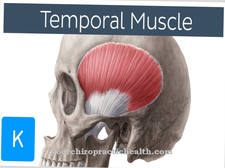
The occiput (os occipitale) and the temporal bone (pars petrosa ossis temporalis) frame the jugular foramen. The petrous bone is part of the temporal bone (os temporale), which in turn is part of the brain skull (neurocranium). The anatomy divides the jugular foramen into either two or three sections.
When divided into three parts, the pars anterior forms the front area of the throttle hole. The inferior petrosal sinus, which is a conduit of blood, runs through this part. The middle part of the jugular foramen is represented by the pars intermedia, which provides access to the brain for the glossopharyngeal nerve, the vagus nerve and the accessory nerve. The core areas of the three cranial nerves are located in the brain stem. The meningeal artery also runs here. Another blood conductor, the sigmoid sinus, runs through the posterior pars. It combines smaller blood streams that divert oxygen-poor blood from the brain, and passes directly at the jugular foramen into the internal jugular artery (vena jugularis interna).
As an alternative to this division into three areas, medicine also uses a dichotomy to describe the jugular foramen. The point of passage of the inferior petrosal sinus and the glossopharyngeal nerve forms the anterior area, while the posterior section contains the posterior meningeal artery, the vagus nerve and the accessory nerve.
Function & tasks
The jugular foramen itself has no active function, but it allows nerve fibers and blood vessels to enter the skull. The sigmoid sinus is a blood vessel that occurs once in each half of the head. Its job is to drain oxygen-depleted blood from the brain.
To do this, it takes blood from other, smaller vessels: the transverse sinus and the superior petrosal sinus and, in some cases, the inferior petrosal sinus. The inferior petrosal sinus contains blood from the middle cranial fossa (fossa cranii media). This has six important openings. At the jugular foramen, the inferior petrosal sinus becomes the internal jugular vein. The posterior meningeal artery also flows through the jugular foramen. It supplies parts of the brain with blood, which contains a lot of oxygen and is therefore of central importance for the survival of brain cells.
The cranial nerves IX – XI run through the jugular foramen. The glossopharyngeal nerve represents the ninth cranial nerve and has six larger branches that innervate the tongue, tonsils and various salivary glands, among other things. In contrast, the vagus nerve or cranial nerve X not only supplies parts of the head and neck, but also areas in the chest and abdomen. The accessory nerve forms the eleventh cranial nerve. Its internal branch runs to the vagus nerve, while the external branch has a motor connection to the sternocleidomastoid muscle and the trapezius muscle.
Diseases
Various neurological syndromes are potentially related to abnormalities in the jugular foramen. Avellis (Longhi) syndrome is associated with damage to the glossopharyngeal and vagus nerves due to lesions in the elongated medulla (medulla oblongata).
Typical symptoms are unilateral paralysis (hemiparesis) on the opposite (contralateral) side and paralysis of the soft palate, vocal cords and throat on the same (ipsilateral) side. Also, Avellis Syndrome can decrease the perception of temperature and pain in the side of the body opposite the damaged side.
Jackson syndrome is also associated with paralysis of the hypoglossal nerve, which can lead to problems with swallowing and speaking, among other things. The reason for this is the impairment of the tongue. Jackson syndrome is one of the oblongata syndromes, as here too the cause of the symptoms lies in the elongated marrow. Other symptoms are also possible. The Sicard syndrome is a neurological disease that is characterized by neuralgia of the glossopharyngeal nerve.
Neuralgia causes sharp or stabbing pain as a result of irritation in the innervation area, but it can spread to other areas. Tapia syndrome affects various cranial nerves; Typical is paralysis of the vagus nerve, nervus accessory and hypoglossal nerve. The tongue, throat, and larynx are paralyzed on the side of the body where the lesion is located. In addition, Tapia syndrome may also affect the sternocleidomastoid muscle and the trapezius muscle.
Villaret syndrome can also be due to damage to the jugular foramen. In this case, characteristic symptoms such as sensory and swallowing disorders and paralysis of the vocal cords, sternocleidomastoid and trapezius muscles are due to nerve damage to the facial nerve, glossopharyngeal nerve, vagus nerve and accessory nerve. The sympathetic nervous system of the neck can also be affected.


