in the Superior cervical ganglion or upper cervical ganglion nerve pathways from the head and neck converge. Anatomically, four broad areas can be distinguished, each of which includes several branches; these rami belong to different nerve tracts and form part of the sympathetic nervous system. Damage to the superior cervical ganglion can lead to the failure of bodily functions.
What is the superior cervical ganglion?
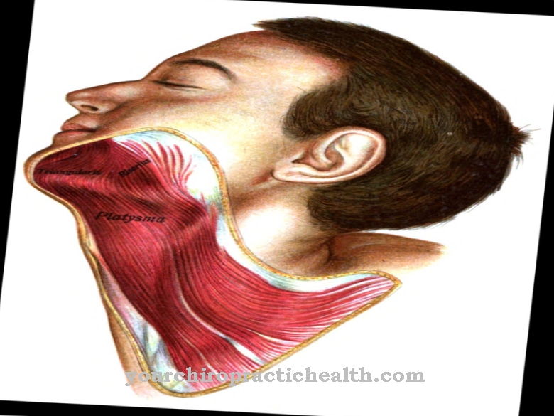
The superior cervical ganglion lies between the longus capitis muscle and the digastricus muscle. The structure at the level of the second cervical vertebra is a collection of nerve cell bodies; these centers form important switching points in the peripheral nervous system and correspond to the basal ganglia or nuclei in the brain.
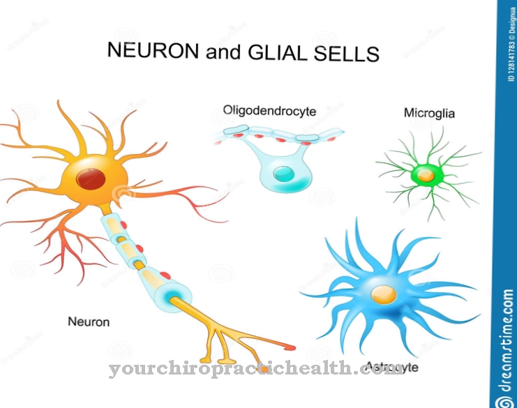
Neuronal cell bodies (somata) lie close together and form connections with one another with their nerve fibers and dendrites. In the cervical ganglion, sympathetic information from the head and neck is combined, which is why the cervical ganglion belongs to the sympathetic trunk (truncus sympathicus). This also includes two further cervical ganglia as well as 20 or 21 other nerve cell body aggregations. Overall, the superior cervical ganglion is 2.5 cm in diameter.
Anatomy & structure
The superior cervical ganglion consists of four areas that can be roughly distinguished without having a clear anatomical barrier. Each of these areas combines several branches, which physiology assigns to different nerves. The anterior branches or rami anteriores form a connection to the head ganglia.
The nerve fibers responsible for this branch further and finally reach the eyes; in addition, they innervate the salivary glands in another branch. The anterior rami of the superior cervical ganglion comprise nerve fibers from the internal carotid nerves and the external carotid nerves. They open into the carotid artery, with the anterior rami winding separately around the inner and outer branches of the blood vessel.
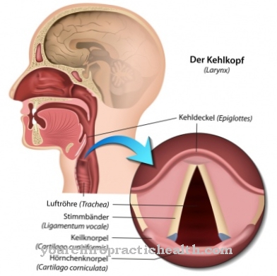
These braids around the carotid artery are called, depending on the location, the internal carotid plexus or external carotid plexus - translated as "braid of the inner carotid artery" or "the outer carotid artery". The medial rami form the middle area of the superior cervical ganglion. They carry nerve signals from / to the heart, larynx and throat. In addition, the upper and middle cervical ganglion (ganglion cervicale medium) are connected via the inferior rami. In contrast, the rami laterales, i.e. H. the lateral branches from the superior cervical ganglion, to the spinal cord, and to various cranial and other nerves.
Function & tasks
The main task of the superior cervical ganglion is to interconnect the nerves from the neck and head area that converge here. Those fibers belong to the sympathetic nervous system, which is a subdivision of the autonomic nervous system. In general, it is considered an activating functional unit. Among other things, it controls the skeletal muscles, the activity of the heart, blood pressure and metabolism in general.
The carotid nerves run from the carotid plexus to the head ganglia and then to the eye and the salivary gland. Neural signals from the nerve fibers trigger the secretion of digestive fluid in the salivary gland. Medicine also knows the organ as the glandula salivatoria and thus describes the entirety of the salivary glands. Three large and five small salivary glands produce secretions for the oral cavity. The jugular nerve also passes through the cervical ganglion.
The rami mediales not only include the sympathetic supply of the larynx and throat, but also contribute to the heart function. The superior cardiac nerve, also known as the superior cardiac nerve, is responsible for this task. In addition to it, there are two other heart nerves: Nervus cardiacus cervicalis medius and inferior. Sympathetic activation accelerates the heartbeat and increases blood pressure. This can be a reaction to physical exertion, stress or fear, for example. In this way, the heart is able to pump more blood and thus ensure the supply of the body under stressful conditions.
Diseases
The ganglion cervicale superius with its interconnections belongs to the vegetative nervous system. For a long time, functions such as heartbeat and blood pressure were considered unaffected; Today, however, recent studies show that patients with high blood pressure can deliberately lower it with adequate exercise.
The training consists of biofeeedback, which visually illustrates the blood pressure and thus gives those affected the opportunity to influence it. Patients who are successful in this cannot control specific muscles, glands or nerves directly, but complex mechanisms allow them to exert an indirect influence. However, this experimental biofeedback approach is still in an early research stage and not every patient is able to achieve an effect. Ancient meditation and trance techniques from Asia may be based on similar biological mechanisms.
In addition to general diseases and nerve damage, two specific clinical pictures can manifest themselves in connection with the superior cervical ganglion. Horner's syndrome manifests itself in a narrowed pupil (miosis), drooping eyelid (ptosis) and an apparent sagging of the eyeball (enophthalmos). Not only lesions on the superior cervical ganglion can trigger Horner's syndrome; Nerve damage in other areas of the sympathetic system can also be considered as a cause.
Familial dysautonomia (Riley Day syndrome), on the other hand, is a genetic disease that leads to the loss of nerve cells. If the superior cervical ganglion is affected, lacrimal fluid may be absent, blood pressure may fluctuate greatly and digestion may be impaired. Other potential symptoms are limitations in temperature perception, gait and speech disorders as well as short stature and curvature of the spine.

