Of the Inferior pharyngis constrictor muscle is the lower throat cords and contributes to speaking and swallowing. Both areas of activity can be disturbed if the inferior constrictor pharyngis muscle fails, is cramped or is otherwise impaired. This is the case, for example, with nerve paralysis or in the context of a peritonsillar abscess.
What is the inferior constrictor pharyngis muscle?
The inferior pharyngeal constrictor muscle is the lower of the three throat cords. The other two muscles in this group are the superior pharyngeal constrictor and the medius pharyngeal constrictor.
Together, the throat constricters transport food or fluid to the esophagus during the pharyngeal transport phase. During pregnancy, the inferior pharyngeal constrictor muscle develops from the sixth gill arch of the embryo. This gill arch also contains the systems for the larynx muscles (Musculi laryngis), parts of the larynx (larynx) and blood vessels.
Since the fourth and sixth gill arches in the embryo merge after a short time, there is a closer spatial and functional relationship between the musculus constrictor pharyngis medius and inferior than between these two muscles and the musculus constrictor pharyngis superior. The latter is located at the top of the throat and, when swallowing, closes the nose together with the soft palate lifter (Musculus levator veli palatini) and the soft palate tensioner (Musculus tensor veli palatini).
Anatomy & structure
The muscle constrictor pharyngis inferior combines two parts: the pars thyropharyngea and the pars cricopharyngea. Both start at the throat suture, which anatomy also calls the pharyngeal raphe. It is located on the back wall of the throat and is partially visible from the outside through the pharynx. The other throat cords also start at the throat seam.
The two parts of the inferior constrictor pharyngis muscle have different origins in the larynx. One of the laryngeal cartilages is the annular cartilago cricoidea, which has grooves. The pars cricoidea of the musculus constrictor pharyngis inferior has its origin at such a notch - the linea obliqua. The pars thyroidea, on the other hand, arises from the cartilago thyroidea, which is also known as thyroid cartilage or thyroid and the pars thyroidea provides support at its outer edge.
Overall, the inferior constrictor pharyngis muscle has a fan-shaped shape. It occurs once on each side of the body and belongs to the striated muscles. Nerve fibers from the ninth and tenth [[cranial nerves (glossopharyngeal nerves and vagus nerves) control the activity of the lower throat constrictor, to which the esophagus is connected.
Function & tasks
The tasks of the inferior constrictor pharyngis muscle include two functional areas. On the one hand it plays a role in speaking and on the other hand it helps swallowing. The inferior constrictor pharyngis muscle influences the position of the larynx over the cartilage where it originates.
At this point, the vocalis and cricothyroideus muscles also act on the vocal folds, which medicine also calls internus and externus. They belong to the muscles of the larynx. In the act of swallowing, the inferior pharyngeal constrictor muscle is active during the pharyngeal transport phase. Before this, the mouth chops up the food in the oral preparation phase and transports the pulp or liquid to the throat in the oral transport phase. The subsequent pharyngeal transport phase consists of a complex interplay of different muscle groups.
The soft palate tensioner (Musculus tensor veli palatini), the soft palate lifter (Musculus levator veli palatini) and the upper throat constrictor (Musculus constrictor pharyngis superior) seal the nasopharynx against penetrating food. With the help of the suprahyoid and infrahyoid muscles, the tongue pushes the contents of the mouth further back into the throat. The musculus constrictor pharyngis medius is responsible for the transport in the oropharynx (mesopharynx), the musculus constrictor pharyngis inferior takes over the transport of the food in the larynx (hypopharynx). The pharyngeal transport phase is followed by the esophageal transport phase, in which the tunica muscularis of the esophagus pushes the bite to the stomach.
You can find your medication here
➔ Medication for tonsillitis & sore throatDiseases
Impairment of the inferior constrictor pharyngis muscle can affect speaking and swallowing. Paralysis of the ninth and tenth cranial nerves, which innervate the lower throat constrictor, are a possible cause of such disorders.
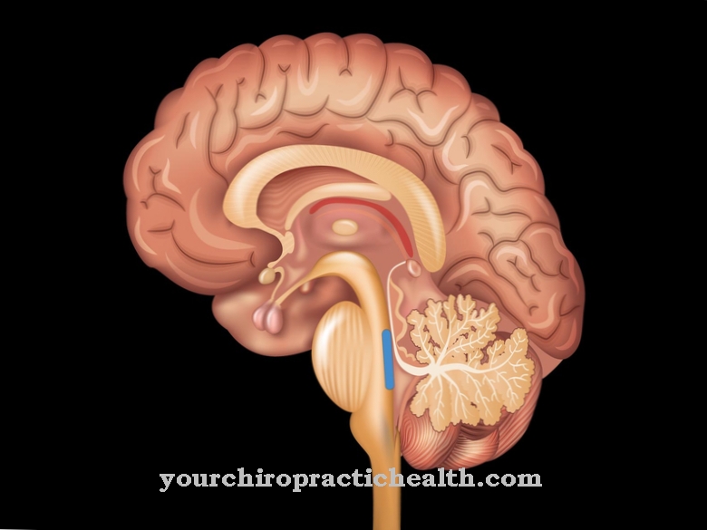
Nerve failure also affects other parts of the speech and swallowing muscles. The fibers of the glossopharyngeal and vagus nerves pass over the pharyngeal plexus. The plexus, like the upper sections of the cranial nerves, can suffer from inflammation, tumors, bleeding, poisoning and injuries. Irradiation of breast cancer rarely leads to unwanted damage to the pharyngeal plexus. Events such as strokes or epileptic seizures and neurodegenerative diseases can also affect the cranial nerves and their core areas in the brain. The extent and duration of the lesion vary greatly from case to case and depend not only on the basic cause, but also on individual factors.
In tonsillitis, the infection sometimes spreads to other tissues. This can also affect the upper almond bay (fossa supratonsillaris), which is connected to the throat constrictions, and cause an abscess. Medicine also calls such pus formation a peritonsillar abscess. This typically causes pain when swallowing, which can radiate to the ear, and leads to swelling in the affected area.
If the masticatory muscles also become inflamed and cramped, those affected also suffer from a clamp (ankylostoma): They can no longer open their mouth unhindered. Other symptoms of a peritonsillar abscess include difficulty speaking and general symptoms such as fever, chills, and malaise.

