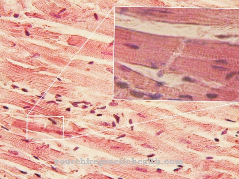
Of the Geniohyoid muscle belongs to the suprahyoid muscles that work together to open the jaw and help swallow.
The hypoglossal nerve is responsible for the nervous supply of the geniohyoid muscle. Accordingly, hypoglossal paralysis impairs the function of the muscle and causes swallowing disorders, as can occur in the course of numerous neurological, muscular and other diseases.
What is the geniohyoid muscle?
One of the suprahyoid muscles in the human jaw region is the geniohyoid muscle, also known as the Chin hyoid muscle is known. The group of suprahyoid muscles includes the geniohyoid muscle, the digastricus muscle, the mylohyoid muscle and the stylohyoid muscle.
These four muscles work together to swallow and open the jaw. The chin hyoid muscle is one of the skeletal muscles that can be deliberately influenced. It is also involved in various reflexes, for example in automated swallowing and breaking. The vomiting center in the brain stem reacts to potentially toxic substances and can trigger the evacuation process. To do this, it coordinates the interaction of various nerves, muscles and glands.
The position of the geniohyoid muscle is a feature that distinguishes modern humans (Homo sapiens) from Neanderthals: the latter had a horizontal chin and hyoid muscle, while the geniohyoid muscle in Homo sapiens is slightly inclined. Perhaps this difference affects the ability to articulate.
Anatomy & structure
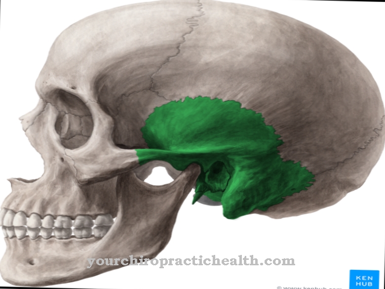
The geniohyoid muscle arises from the mental spine, which forms a protrusion in the lower jaw bone (os mandibulare) and can be found there on the inner surface (facies interna). The insertion of the muscle is on the hyoid bone (os hyoideum).
In fine construction, the geniohyoideus muscle consists of striated muscle tissue, the name of which goes back to the easily recognizable fiber structure. The individual elongated muscle fibers are each surrounded by a layer of connective tissue; inside are the thread-like myofibrils. The sarcoplasmic reticulum, which corresponds to the endoplasmic reticulum of other cells, wraps around them. The myofibrils can be divided into transverse sections known as sarcomeres. A Z-disk delimits the sarcomere on both sides and serves as a hold for tiny filaments.
According to the zipper principle, filaments made of actin and tropomyosin on the one hand and myosin on the other hand are arranged alternately so that they can slide into one another when the muscle contracts. The geniohyoid muscle receives such neuronal signals via the hyoglossal nerve, which is connected to the spinal cord via the spinal segment C1 and also innervates the other suprahyoid muscles.
Function & tasks
The function of the geniohyoid muscle is to assist in opening the jaw and swallowing, pulling the tongue forward. It is also involved in sideways movements of the jaw and, together with the other suprahyoid muscles, forms the muscles of the floor of the mouth. Motor fibers of the hypoglossal nerve transmit signals to the geniohyoid muscle by releasing neurotransmitters at the junction between the nerve fiber and the muscle cell.
These messenger substances are reversibly attached to receptors located on the outside of the muscle cell membrane. An activated receptor opens ion channels through which charged particles flow into the cell and create an electrical endplate potential in the muscle. This spreads over the tissue of the geniohyoid muscle and stimulates the sarcoplasmic reticulum to release calcium ions.
The ions bind to the actin / tropomyosin filaments of the fine myofibrils, which are bundled in the muscle fiber, and in this way change their spatial structure. As a result, the myosin filaments with their “heads” find a hold on the actin / tropomyosin strand. The myosin filaments push themselves further between the complementary fibers and thereby actively shorten the sarcomere and ultimately the entire muscle. The contraction of the geniohyoid muscle pulls the tongue forward.
You can find your medication here
➔ Medicines for sore throats and difficulty swallowingDiseases
A lesion on the hypoglossal nerve can impair the function of the geniohyoid muscle if the innervating fibers no longer transmit nerve signals to the muscle. Typically, hypoglossal paralysis affects not only the geniohyoid muscle, but also the other muscles of the tongue.
The nerve is often damaged in only one half of the face, which results in paralysis of the tongue on one side. At the functional level, this paralysis often leads to swallowing disorders (dysphagia) and motor problems when speaking. The position of the tongue often deviates from its normal position in the mouth. Persistent hypoglossal paralysis gradually leads to the atrophy of the affected muscles, which leads to the easily recognizable asymmetry, which is particularly noticeable when the tongue is stuck out.
Various causes can be considered for hypoglossal paralysis, including a stroke or a cerebral infarction. In Germany, 160–240 of every 100,000 people suffer an ischemic stroke, which is the most common form of cerebral infarction and is due to the insufficient supply of blood to the brain. Symptoms can vary depending on the area affected. Hypoglossal paralysis can also be permanent if the nerve tissue is permanently damaged.
Swallowing disorders can also appear, particularly in the advanced course of Alzheimer's disease. The neurodegenerative disease shows itself at the beginning in disorders of the short-term memory and leads to increasing symptoms such as agnosia, apraxia, language and speech disorders, apathy and ultimately to bed rest and numerous motor disorders. In addition to malformations and neoplasms, neuromuscular diseases are other possible causes of swallowing disorders that involve the geniohyoid muscle and other muscles. Direct injuries to the geniohyoid muscle are possible when using implants and other injuries and fractures in the facial area.

