As external tongue muscle is the Hyoglossus muscle involved in swallowing, speaking, sucking and chewing, pulling the tongue back and down. Functional limitations are often due to problems with the hypoglossal nerve, which supplies the muscle neuronally.
What is the hyoglossus muscle?
The hyoglossus muscle is one of four outer muscles of the tongue, which also includes the genioglossus muscle, the styloglossus muscle and the chondroglossus muscle.
Because of its location in the body, the hyoglossus muscle is also known as the hyoid-tongue muscle. The contraction of the muscle causes the tongue to move back and down. Its antagonist is the styloglossus muscle, which is another external tongue muscle and is primarily involved in swallowing. When it contracts, it pulls the tongue back and up, partially relaxing the hyoglossus muscle.
Experts do not agree on whether the chondroglossus muscle belongs to the hyoglossus muscle and splits off from it - or whether it is a separate muscle. The chondroglossus muscle is two inches long and pulls the tongue back and down like the hyoglossus muscle. It arises from the hyoid bone and attaches to the tongue.
Anatomy & structure
The origin of the hyoglossus muscle is in the lower rear area of the oral cavity on the hyoid bone (os hyoideum). The hyoid bone is a bone held by muscles and ligaments without being directly connected to other bones - but the hyoglossus muscle is not one of its supporting muscles.
Instead, he in turn depends on the firm hold of the hyoid bone. The insertion of the musculus hyoglossus is attached to the aponeurosis linguae. The tendon plate is located between the tongue muscles and the oral mucosa and merges into the tongue septum (septum linguae) with which it is fused. In its basic form, the hyoglossus muscle forms an approximately square, thin surface. It belongs to the striated skeletal muscles, the structure of which consists of individual fibers.
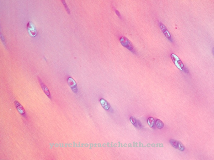
Such a muscle fiber or muscle cell arises from cell division and has many cell nuclei, which, however, are not, as usual, located in a separate cell. Instead, they form a fabric with a superordinate organization. A muscle fiber combines many myofibrils. The striated muscles owe their name to their microscopic image: alternating light and dark stripes. They come about because hair-like fibers made of actin and myosin are shifted closer or further into one another.
Function & tasks
The hyoglossus muscle participates in swallowing, speaking, sucking and chewing. The cranial nerve XII or the hypoglossal nerve, which also innervates the other tongue muscles, is responsible for its control. The nerve carries commands to tense the muscles in the form of electrical impulses that travel along the nerve fiber.
At the muscle, the fiber ends in a motor end plate: it contains vesicles that are filled with neurotransmitters. The incoming electrical stimulus leads to the release of the transmitter into the synaptic gap between nerve and muscle. When they reach the membrane of the muscle cell, the molecules open ion channels, which slightly changes the charge state of the cell. This temporary electrical charge on the muscle cell is also known as the endplate potential. It migrates via the sarcolemma and T-tubules to the sarcoplasmic reticulum, which then releases calcium ions.
Calcium binds to the fine structures of the myofibrils and ensures that its actin and myosin filaments slide into one another. This causes the irritated muscle fibers to shorten lengthways and pull the tongue backwards and downwards at the same time, which is necessary when swallowing, speaking, sucking and chewing. Humans are able to consciously control these movements; however, automatic reflexes also have an influence on the control of the hyoglossus muscle. For example, the sucking reflex in newborns is not the result of an arbitrary action, but part of an innate behavioral program.
You can find your medication here
➔ Medicines against loss of appetiteDiseases
Since the hyoglossus muscle is located well inside the head, direct lesions of the tissue are rare. Functional failures and complaints of the tongue muscle are often due to damage to the hypoglossal nerve, which is responsible for its control.
Medicine differentiates between unilateral and bilateral lesions, both of which lead to various disorders of chewing, swallowing, sucking and speaking. The causative lesion of the hypoglossal nerve can itself be due to injuries, neurodegenerative diseases or a stroke.
A bilateral lesion is reflected in the complete paralysis of the tongue: The tongue is completely inoperable because the hypoglossal nerve not only innervates the hyoglossus muscle, but is also responsible for controlling the other tongue muscles. If the nerve damage persists, the muscle tissue disappears (atrophy) as the body gradually breaks it down. If it is a reversible lesion on the hypoglossal nerve, training of the affected muscles is often necessary after the paralysis of the tongue. Targeted exercises stimulate the body to rebuild the tissue. The extent to which a complete restoration of the normal state is possible depends on the individual case.
In contrast to complete paralysis of the tongue, unilateral paralysis of the tongue is due to a unilateral lesion on the hypoglossal nerve. As a result, the tongue hangs down on the affected side. Conversely, a slight deviation in the position of the tongue does not necessarily indicate nerve damage, as it can also be due to other factors and is not always pathological.






.jpg)


.jpg)




.jpg)
