Of the Masseter muscle is one of four muscles of mastication. The skeletal muscle is involved in the crushing of the food and in the salivation of the food pulp. The masticatory muscle can be affected by pathological tension up to locking the jaw as well as inflammation and paralysis.
What is the masseter muscle?
The skeletal muscles are largely made up of muscles with skeletal fixation. Skeletal muscles, including their neuromuscular components, are essentially involved in every movement of the body and are part of the striated muscles. The skeletal muscles include the masticatory muscles.
This part of the skeletal muscles is made up of four different muscles that attach to the mandible and are involved in the act of chewing. One of these four muscles is the masseter muscle, which is formed during embryonic development together with the other masseter muscles from the connective tissue parts of the first gill arch. The muscle is involved in what is known as the masseter reflex. This is a self-reflex of the masticatory muscles, which is preceded by a blow to the lower jaw. The masseter reflex is one of the innate protective reflexes of the human jaw. Basically all mammals have a masseter muscle. The exact anatomy of the muscle differs from species to species.
Anatomy & structure
In humans, the masseter muscle arises on the zygomatic arch. For other mammals, the skeletal muscle often arises from the facial crest on the upper jaw. The human masseter muscle is a feathered muscle that macroscopically consists of a superficial and a deep part.
The superficial part runs obliquely in the dorsal-caudal direction and thus reaches the mandibular branch and the masseteric tuberosity. The deep part of the muscle runs in the vertical in a caudal direction and thus extends to the ramus mandibulae. The masticatory muscle is motorized by the masseteric nerve, a branch of the mandibular nerve. The arteria transversa faciei is responsible for the blood supply to the muscle in addition to the arteria masseterica. In humans, a parotid duct runs through the skeletal muscle. The masseter muscle, like the other three masseter muscles, is arranged so that it can be moved to a large extent. For this purpose, firm fasciae envelop the masseter muscles.
Function & tasks
The masseter muscle, together with the temporalis and medial pterygoid muscles, close the jaw. In this way, the muscle allows the actual jaw closure on the one hand and lateral and longitudinal movement of the lower jaw on the other. During the act of chewing, the movements of the muscles are involved in the crushing of the food and thus partially ensure food intake. In addition, the muculus masseter triggers a chain reaction that is just as important for food intake.
The movements of the muscle massage the parotid gland as part of the act of chewing. The gland is the paired parotid gland, whose job it is to produce saliva. By stimulating the gland, saliva is secreted. As a result of the chewing movements, the saliva produced reaches solitary single glands in the pharynx and oral mucosa via the ducts. In this way, by stimulating the parotid gland, the masseter muscle ensures that the chewed food is penetrated with saliva. The salivary enzymes initiate the digestion process of sugar molecules such as starch and break down proteins using proteases.
The food pulp produced by the chewing movement is prepared for digestion in the stomach. In addition, the penetration of the pulp with saliva facilitates the swallowing process. Apart from these tasks, the muculus masseter fulfills protective functions for the jaw as part of the masseter reflex. The reflex movement corresponds to a muscle stretching reflex and is one of the protective reflexes of the jaw. When the skeletal muscle is stretched in length by a blow to the lower jaw, the masseter reflex, like any other muscle stretching reflex, causes the muscle to contract. The jaw closes by connecting loops of afferent and efferent neurons.
Diseases
The masseter reflex in particular is part of the neurological reflex examination. The examiner checks with light strokes on the lower jaw whether the innate masseter reflex is preserved in the patient. Abnormal reflex responses can imply trigeminal nerve paralysis. Lesions of the trigeminal nerve trigger peripheral paralysis, such as those that occur in the context of polyneuropathy.
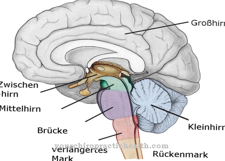
This disease is mostly due to causes such as malnutrition, poisoning, infections or trauma. Central nerve lesions can also be indicated by an altered masseter reflex, especially those in the area of the brain stem. In such a case, tumors, strokes and inflammatory processes are possible causes. In addition to neuromuscular paralysis, the masseter muscle can gain pathological relevance in the context of pain symptoms. Pain within the masticatory muscles is around three times as common as temporomandibular joint pain. This myofascial pain can radiate over the entire neck and back area and is often the result of incorrect bites and repeated incorrect strain on the masticatory muscles.
Under certain conditions, the masseter muscle can become painfully inflamed. This kind of inflammation can result from bacterial infection. Much more often, however, inflammation of the skeletal muscle occurs in the context of muscle overload or missed bites. In addition to the phenomena mentioned, pathological phenomena such as the lock jaw are associated with the jaw muscles. The cause of a locked jaw is a spasm of the masticatory muscles.
The masseter muscle is also sometimes affected by tension. Tensions of this kind are particularly noticeable during the act of chewing in the form of pain in the jaw. Atrophy of the masseter muscle occurs less frequently than the phenomena mentioned. With this phenomenon, the masseter gradually decreases in mass due to interrelationships such as immobilization.


