Of the Stylohyoid muscle is a small skeletal muscle in the jaw area. It is part of the suprahyoid muscles and helps swallow and open the jaw. Swallowing disorders (dysphagia) can also affect the stylohyoid muscle and lead to functional impairments.
What is the stylohyoid muscle?
The stylohyoid muscle is a striated muscle that participates in opening the jaw and swallowing. It belongs to the group of suprahyoid muscles, also known as the floor of the mouth muscles or the upper hyoid muscles, and includes four other muscles in addition to the stylohyoid muscle: the digastric muscle, the geniohyoid muscle and the mylohyoid muscle.
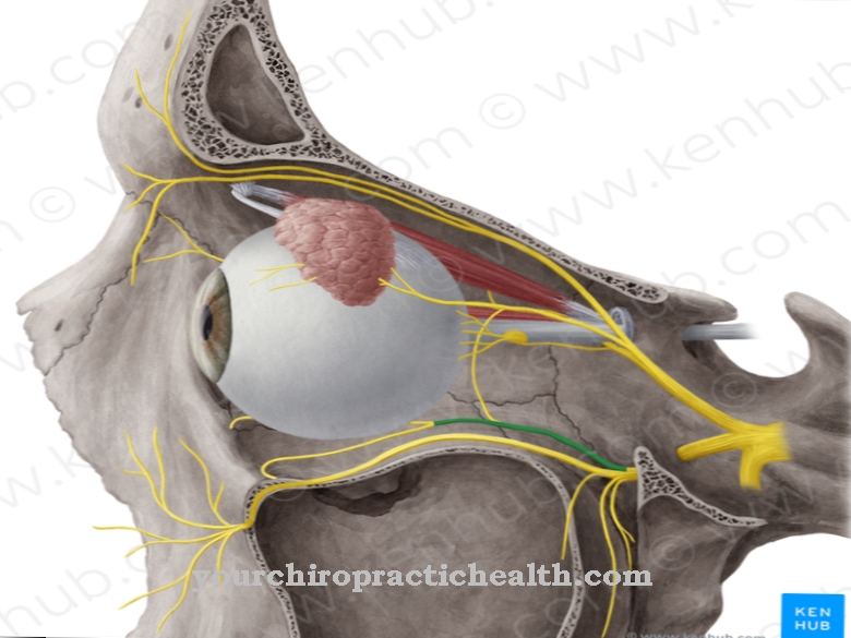
Both when swallowing and when opening the jaw, these muscles work together in a coordinated manner. The control is based on the seventh cranial nerve, which penetrates as the facial nerve or the facial nerve with the help of numerous branches (rami) to numerous tissue structures in the head. Its fibers not only conduct motor and parasympathetic signals from the central nervous system to the innervated muscles, but they also transport sensory and sensitive nerve signals in the opposite direction.
Anatomy & structure
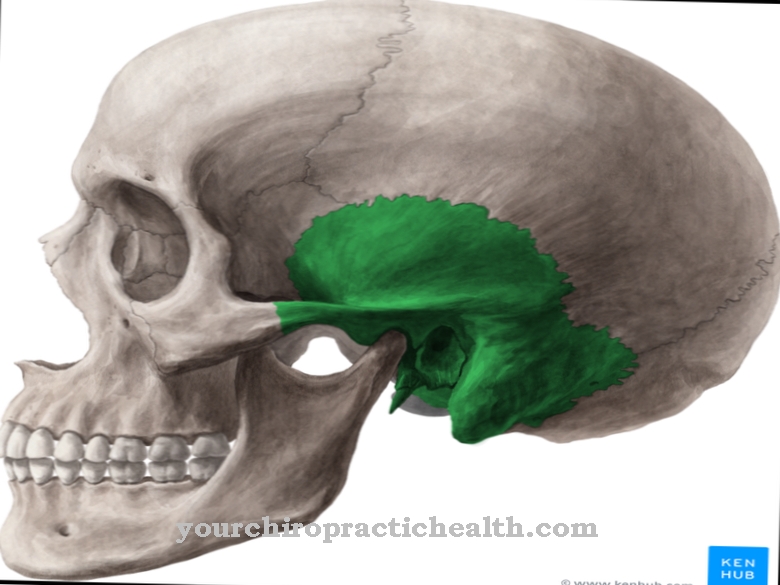
The stylohyoid muscle originates from the temporal bone (os temporale), which is part of the skull. The inner ear and middle ear lie in it. On the temporal bone, the stylohyoid muscle arises from the stylus extension (processus styloideus), which is an extension of this skull bone.
The insertion of the stylohyoid muscle is located on the hyoid bone (os hyoideum), where a tendon fixes the striated muscle to the bone and also attaches the digastric muscle tendon. The digastric muscle is another suprahyoid muscle that is also known as a dibular muscle due to its shape. The stylohyoid ligament - a pair of ligaments - extends from the stylus process to the hyoid bone and connects the two bones with one another.
Like all striated skeletal muscles, the stylohyoid muscle is made up of muscle fibers that correspond to muscle cells. They have several cell nuclei because the conventional cell structure does not exist in them. Instead, there are several myofibrils inside a muscle fiber, which run lengthways through the fiber and are surrounded by the sarcoplasmic reticulum. When the transverse sections of the myofibrils (sarcomeres) shorten because the actin / tropomyosin and myosin filaments they contain slide into one another, the muscle as a whole contracts, causing the hyoid bone to move accordingly.
Function & tasks
The stylohyoid muscle has both a static and a dynamic function. Together with other muscles and ligaments, it holds the hyoid bone (hyoid bone), which otherwise has no direct connection to other bones. The hyoid bone is composed of the middle body and the lateral horns; the insertion of the stylohyoid muscle is distributed over the body and the large horn of the bone.
The dynamic function of the stylohyoid muscle is to assist in swallowing and jaw opening, working in conjunction with the other suprahyoid muscles. The stylohyoid muscle receives the command to contract from the facial nerve. The electrical signal ends in the terminal button of the innervating nerve fibers and is accompanied by an influx of calcium ions. As a result, some vesicles that are located in the terminal button combine with the outer membrane and release the neurotransmitters contained in them.
As a messenger substance, acetylcholine binds temporarily to receptors in the membrane of a muscle cell and causes the influx of ions there, which generate a new electrical potential: the endplate potential, which is transferred to the sarcoplasmic reticulum via sarcolemma and tubular T-tubules. Calcium ions from the sarcoplasmic reticulum penetrate into the interior of the myofibrils and bind to the filaments there, which then slide into one another. In this way, the muscle fibers of the stylohyoid muscle shorten and pull the hyoid bone backwards and upwards, for example when swallowing. In addition to the suprahyoid muscles, the infrahyoid muscles (lower hyoid muscles) also participate in this process.
You can find your medication here
➔ Medicines for sore throats and difficulty swallowingDiseases
Since the facial nerve connects the stylohyoid muscle to the nervous system, damage to the facial nerve can also affect the stylohyoid muscle. Swallowing disorders are summarized in medicine under the term dysphagia.
One of the possible causes is Alzheimer's dementia, which is characterized by progressive damage to the brain, which leads to functional impairments or failures in the affected areas. Parkinson's disease, which is based on the loss of nerves in the substantia nigra, or a stroke, the hereditary disease Huntington's disease or other neurological diseases are possible causes of swallowing disorders. Injuries to the tongue and fractures to the midface or hyoid bone can damage both the muscles and the innervating nerve fibers.
Malformations and neoplasms in the head, diseases of the esophagus and infectious diseases can also contribute to swallowing disorders, which are reflected in functional disorders of the stylohyoid muscle and other muscles involved. Mentally caused swallowing difficulties occur, for example, in the context of phagophobia, which represents a disease-related fear of suffocation or swallowing and is also known colloquially as fear of swallowing.
Eagle syndrome also manifests itself in the vicinity of the stylohyoid muscle. Watt Weems Eagle was the first to describe the disease; it does not affect the stylohyoid muscle directly, but rather the stylohyoid ligament. In Eagle syndrome, calcium salts accumulate in the ligament and cause ossification. The syndrome can also be due to the fact that the stylus process is too long. In both cases, swallowing difficulties such as pain in the throat and difficulty swallowing when turning the head are typically evident.



.jpg)









.jpg)