At a Muscle biopsy Doctors extract muscle tissue from the skeletal muscles to diagnose neuromuscular diseases, for example in the presence of myopathies. Another task of the muscle biopsy is to examine the preserved tissue material. Closely related areas of expertise are neurology, neuropathology and pathology.
What is the muscle biopsy?

Various disease processes can cause pain or muscle weakness. These abnormalities lead to permanent problems and diseases of the connective tissue, nervous system, vascular system or musculoskeletal system. In the field of sports medicine, muscle biopsies are performed to gain knowledge about muscle metabolism during and after physical exertion.
The muscle biopsy is induced in the case of atypical or unusual complaints or if symptoms are predominantly limited to the muscles near the trunk (proximal). Tissue extraction is an important medical instrument for differential diagnostic findings when ALS (degenerate disease of the motor nervous system) is suspected. However, it is not necessary in every case. The findings regarding changes in muscle tissue, especially in diseases of the second motor neuron, are based on the evaluation of frozen muscle sections, which are routinely stained and examined for the existence of special enzymes using certain reagents. In ALS, only slightly weakened muscles are selected for the biopsy.
As a rule, the four-headed thigh muscle (quadriceps muscle), the anterior lower leg muscle (tibialis anterior muscle) or the upper arm flexor (biceps muscle) are used for a biopsy. Muscles that have been damaged by unspecific effects such as direct trauma, entrapment of a nerve or a nerve root lesion are unsuitable. A muscle that is injured, has been the subject of EMGs in the past three weeks, or has recently received frequent injections is unsuitable for the biopsy.
Function, effect & goals
The aim of the muscle biopsy is to ensure that appropriate treatment is initiated after diagnosis. It enables doctors to identify abnormalities in the musculoskeletal system being examined. A muscle biopsy is straightforward and is performed under local anesthesia. For this process, the doctor selects a clearly diseased muscle that is not yet completely fatty or atrophic.
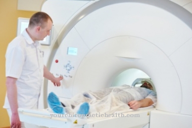
The clinical aspect or the results of examinations carried out (sonography, magnetic resonance tomography) form the basis for the selection of the appropriate muscle. If the choice of tissue cannot be conclusively clarified, electromyography (EMG) or an MRI are used. To avoid incorrect results, the biopsy is not performed in areas where EMG electrodes have been placed or intramuscular injections have been made because the muscle tissue is damaged. There are two types of biopsy: the open biopsy and the punch biopsy. Open tissue collection is the standard procedure. The local anesthetic is not injected into the directly affected tissue, but into the adjacent skin structures.
Then a small incision is made to expose the affected muscle. A tissue sample is taken from this and the wound is closed with a suture after hemostasis. A punch biopsy takes tissue using a biopsy needle that is inserted percutaneously (under the skin) into the muscle. This tissue collection is less invasive than the open method, but only a very small sample can be obtained.
If there is a suspicion of connective tissue diseases of the vessels, areas of the surrounding skin, fascia and subcutaneous fatty tissue are removed in addition to the muscles. The biopsate obtained is further processed in a pathological institute. Preferably, a 2 to 3 centimeter long and 0.3 to 0.5 centimeter thick muscle bundle is attached to a rod (sterile cotton swab) at two ends in the direction of the muscle fibers in order to maintain the orientation of the tissue fibers Rods excised and immediately fixed.
A buffered six percent glutaraldehyde solution, consisting of 20 to 30 millimeters with phosphate buffer, is suitable as a means of fixation for the electron microscopic examination and the semi-thin section method. A similar preparation, fixed in a four percent formaldehyde solution in paraffin embedding, is suitable for examination with a light microscope. Subsequently, an approximately 1 x 0.5 x 0.5 cm section of muscle is excised for immunohistochemical, enzyme-histochemical and molecular biological examination. This piece cannot be fixed or tied to a stick, but must be frozen immediately in liquid nitrogen or immediately taken to the pathology department in a closed vessel with a damp cloth to prevent it from drying out.
The pathologists take care of the processing and carry out the histological examination. Due to the limited shelf life, the items are shipped by courier. The glutaraldehyde and formalin-fixed samples are sent in separately from the frozen muscle section. The containers with the muscle sections placed in the fixation solutions are attached to the outside of the styrofoam box using adhesive strips. If they are in close proximity to the dry ice, the solutions freeze and serious artifacts occur.
Tissue extraction is induced in the following diseases:
- Inflammation of the muscles (polymyositis, inclusion body myositis)
- systemic inflammatory diseases (vasculitis, eosinophilic syndromes)
- Muscular dystrophies (girdle dystrophy, Duchenne muscular dystrophy,)
- Congenital myopathies (nemalin myopathy, central core myopathy)
- neurogenic muscular atrophies (amyotrophic lateral sclerosis, spinal muscular atrophies)
- Myopathies in metabolic disorders (lipid storage myopathies)
- mitochondrial diseases (myoclonus epilepsy with "ragged red" fibers)
- toxic myopathies (chloroquine, colchicine, statins)
- Rhabdomyolysis, muscular dystrophy (muscle wasting)
- unclear diseases of the muscles
Routine pathological examinations are:
- Elastika van Gieson staining (EvG) (fibrosis of the endomysial connective tissue in myopathies)
- Modified Gömöri trichrome staining (inclusion bodies in nemalin myopathy)
- Hematoxylin-eosin stain (inflammatory infiltrates in myositis)
- Oil red color (lipid storage in the case of deficiency symptoms of carnitine palmitoyl transferase)
- Acid phosphatase reaction (increased macrophage activity in inflammatory myopathies)
- ATPase reaction at different pH values (different fiber types and their disturbed distribution in the case of chronic neurogenic damage)
- NADH reaction (illustration of the oxidative, intermyofibrillary network and its disorders in multicore myopathy, central core myopathy)
- PAS staining (increased storage of glycogen in McArdle's disease)
You can find your medication here
➔ Medicines for muscle weaknessRisks, side effects & dangers
Rare complications are infections and wound healing disorders. Since skeletal muscle tissue is maximally irritable and prone to artifacts, there is a risk of crushing or further injury to the tissue. Bruising, discomfort and light bleeding at the donor site are possible. Before the procedure, the doctor informs the patient about the individual risks and asks about contraindications, for example allergies to the anesthetics used. Bleeding disorders, aspirin and anticoagulants (drugs to thin the blood) are important contraindications that may only allow surgery if the drug is discontinued.
To ensure that the patient is physically fit for the procedure, the medical professional will conduct a physical examination in addition to taking the medical history. After the procedure, the patient can quickly resume their normal everyday life, there are only minor restrictions. He must keep the interface sterile and dry and must not put too much strain on the affected muscle tissue.
Typical & common muscle diseases
- Torn hamstring
- Muscle weakness
- Compartment syndrome
- Inflammation of the muscles (myositis)
- Muscle wasting (muscular dystrophy)










-eisenmangelanmie.jpg)

.jpg)

