The Odontoblasts are tooth-forming mesenchymal cells of the dentition and secrete so-called predentin to dentinize the teeth. After the teeth have formed, they maintain the teeth and repair them in the event of chewing and tooth decay. In the case of avitaminoses such as vitamin C deficiency, irreversible degeneration of the cells often occurs.
What are Odontoblasts?
With the milk teeth and the change of teeth, two tooth formation processes take place in the human organism. The odontoblasts play an important role in these processes. These are highly specialized cells in tooth tissue.
They are of mesenchymal origin and develop from the ectodermal neural crest. After their differentiation, the cells play a key role in the formation of teeth. They produce predentin, which is known as the organic precursor of dentin, for a lifetime. In terms of tooth development, the formation of dentin is referred to as dentinization or dentinogenesis. Odontoblasts provide the necessary material for this dentinogenesis.
As cells of the mesenchymal connective tissue, they are related to the osteoblasts and fibroblasts. Just as osteoblasts take on bone-building tasks, they also have tooth-forming functions. Apart from the hard enamel, the mesenchyme provides all components of the teeth. Due to their direct connection to the nervous system, odontoblasts also play a decisive role in the sensation of pain in the teeth.
Anatomy & structure
During tooth formation, the epithelial cells in Hertwig's vagina initiate the formation of osteoblasts. They cause the cells of the adjacent mesenchyme to differentiate. This is how odontoblasts develop from the mesenchymal cells.
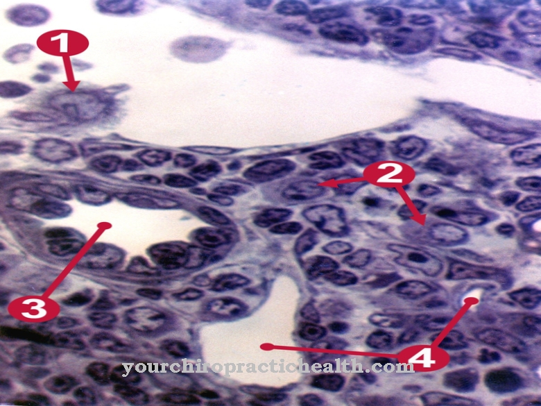
The odontoblasts then find themselves in the border area between pulp and dentin. The former mesenchymal cells have a cylindrical shape and a palisade-like arrangement. Because they form dentin throughout their life, the pulp cavity decreases in size with increasing age. The fine cell processes of the odontoblasts are called Tomes fibers. During the formation of dentine, the predentine becomes calcified on these structures, creating dentine tubules. These canals are called Tomes canals and correspond to the fine, hair-like cavities that run through the dentin from the inside out.
The canals are filled by the up to five millimeter long processes of the odontoblasts. Each odontoblast is also in direct contact with free nerve endings.
Function & tasks
Odontoblasts secrete predentin, which is known as the organic precursor of dentin, to form teeth. They are thus significantly involved in odontogenesis. Dentin formation is also known as dentinogenesis. In the course of tooth formation, this process appears as the first recognizable feature of the crown stage. The odontoblasts differentiate themselves from the dental papilla cells and secrete an organic matrix at the later tooth tip, which is close to the inner epithelium.
The matrix consists of collagen fibers with a diameter of up to 0.2 μm. The odontoblasts migrate to the center of the future tooth. There they form offshoots, which is also called the odontoblast process. The offshoot initiates the secretion of hydroxyapatite crystals. The mineralization of the organic matrix begins. The mantle dentine is formed from already existing basic substances of the dental papilla. Primary dentin is created by processes of the odontoblasts. The cells grow in size until extracellular resources can no longer contribute to the organic matrix. Large odontoblasts secrete little collagen and allow structured heterogeneous nuclei to grow.
In addition to the collagen secretion, lipids, phosphoproteins and phospholipids are secreted at this stage. When the tooth formation is complete, the odontoblasts lose their ability to divide. They come to rest in the pulp periphery and maintain the dentin coat of the teeth for the rest of their life by adding secondary and tertiary dentin. Secondary dentin forms significantly more slowly than primary dentin. The formation only takes place after the root formation has been completed. In the immediate vicinity of the crown, development is faster than elsewhere on the tooth. Tertiary dentine is also known as repairing dentine and is reactive to chewing or caries.
You can find your medication here
➔ Medication for toothacheDiseases
Vitamin deficiency can affect the odontoblasts. This is especially true for vitamin C deficiencies. Avitaminosis C is also known as scurvy and used to be common among sea travelers without a balanced range of foods.
The associated deficiency in ascorbic acid endangers the cohesion of the tissue, since sufficient cement substance can no longer be produced. Small-sized bleeding occurs in the muscles through capillary blood escapes. Cartilage cells and epiphyses become detached in the bones, and edema often develops in the mouth. A deficiency in vitamin C hits the odontoblasts just as badly. They slowly degenerate and no longer give off enough dentin. They are closed off by the predent, which further favors their degeneration. Since the degenerated cells are no longer able to repair the teeth due to the reduced dentin production, the teeth are hit all the harder by diseases such as caries.
Dentin dysplasia of the radicular and coronal forms or dentinogenesis imperfecta are somewhat less common than avitaminoses. In these hereditary diseases, dentinogenesis is disturbed by the odontoblasts. Large cavities show up within the dentin. The teeth wear out more easily and are more vulnerable to fractures. The symptoms of hereditary diseases can be alleviated by endodontological and endosurgical measures as needed. If the teeth cannot be preserved, they are removed. After the removal, an implantation can take place if necessary.
Typical & common dental diseases
- Tooth loss
- Tartar
- Toothache
- Yellow teeth (tooth discoloration)








.jpg)

.jpg)
