They are in the midbrain Pedunculi cerebri, which are composed of the cerebral legs (crura cerebri) and the midbrain dome (tegmentum mesencephali).
Lesions in these areas can be associated with various complaints, depending on which structures are affected. For example, Parkinson's disease is due to a decrease in the substantia nigra in the tegmentum and typically leads to tremor, rigidity, bradykinesia and postural instability.
What are the pedunculi cerebri?
The pedunculi cerebri are also called (Large-)Brainstalks known and are located in the midbrain (mesencephalon). In the two halves of the brain, they form a symmetrical structure and can be anatomically divided into cerebral crura (crura cerebri) and midbrain hood (tegmentum mesencephali), with the tegmentum having a significantly larger proportion.
More rarely, the specialist literature equates the pedunculi cerebri with the crura cerebri without including the tegmentum. The midbrain consists not only of the crura cerebri and tegmentum mesencephali, but also has a third part: the roof of the midbrain or tectum mesencephali. The mesencephalic aqueduct separates the tectum from the tegmentum and is filled with fluid.
Anatomy & structure
The pedunculi cerebri are located in the midbrain and are composed of the crura cerebri and the tegmentum mesencephali. The crura cerebri are divided into a right and a left area; the pit between these two parts is the interpeduncular fossa.
Nerve fibers from the internal capsule run through the crura cerebri and connect the cerebral cortex with deeper brain areas. The internal capsule unites different nerves of the brain and thus represents an important data pathway in the human brain.
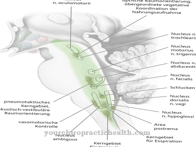
The substantia nigra, which belongs to the extrapyramidal motor system and lies next to the crura cerebri, belongs to the tegmentum mesencephali. In addition, the tegmentum mesencephali contains parts of the formatio reticularis and other core areas that consist of gray matter: the density of cell bodies in them is particularly high.
These core areas form the origin of cranial nerves, which run from them through the brain. Nerve pathways can carry both afferent fibers, which transport information from the peripheral nervous system to the brain, and efferent fibers, which transmit signals from the central nervous system to the periphery. The nuclei of the tegmentum include:
- Nucleus ruber
- Nucleus nervi oculomotorii
- Nucleus accessorius nervi oculomotorii
- Nucleus nervi trochlearis
- Nucleus mesencephalicus nervi trigemini
Function & tasks
Although the pedunculi cerebri anatomically form a coherent complex, their tasks differ depending on the area. Numerous nerve tracts run through the internal capsule in the cerebral crura, and three parts of the inner capsule can be distinguished. The anterior crus is formed by the nerve tracts that run between the caudate nucleus and the putamen / pallidum. These include the thalamic stem and the frontopontinus tract, which forwards information from the frontal lobe to the pons.
The posterior crus of the internal capsule includes the pyramidal tract, which is responsible for controlling movement, and fibers of the auditory and visual tract. Between the crus anterius and the crus posterius lies the genu ("knee") of the inner capsule.
The substantia nigra and the nucleus ruber, which belong to the extrapyramidal motor system, are located in the tegmentum mesencephali. The fibers of the third cranial nerve originate from the nucleus nervi oculomotorii and the nucleus accessory nervi oculomotorii. This controls movements of the eyes and is also known as the oculomotor nerve. Its three branches are the superior ramus, the inferior ramus and the ramus to the ciliary ganglion. The latter is a collection of nerve cell bodies in the peripheral nervous system that is located in the eye socket.
Another cranial nerve is the trochlear nerve. Its origin also lies in the tegmentum mesencephali of the pedunculi cerebri: the nucleus nervi trochlearis is responsible for the nerve tract. As the fourth cranial nerve, the trochlear nerve is also responsible for eye movements. In contrast, the nucleus mesencephalicus nervi trigemini is a sensitive nucleus and belongs to the nervus trigeminus (cranial nerve V). The nucleus mesencephalicus nervi trigemini processes proprioceptive perceptions from the external eye muscles, the masticatory muscles, the temporomandibular joint and the tooth support system (paradontium).
You can find your medication here
➔ Medicines to calm down and strengthen nervesDiseases
Due to the different structures that lie in the pedunculi cerebri, numerous ailments and diseases are possible in connection with the brain stalks. The substantia nigra lies in the tegmentum mesencephali; in Parkinson's disease the substance disappears and causes a dopamine deficiency in the brain.
Dopamine is an important messenger substance and is of great importance for the transmission of information in the extrapyramidal motor system from one nerve cell to another. The typical symptoms of Parkinson's disease therefore include muscle tremors (tremor), muscle rigidity (rigidity), slowing down of movements (bradykinesia) and postural instability (postural instability). To compensate for the lack of dopamine, doctors can use the active ingredient L-Dopa. This is a dopamine precursor that can cross the blood-brain stage. Other therapy options include catechol-O-methyltransferase inhibitors (COMT inhibitors), which can slow down the breakdown of dopamine or L-dopa, or targeted stimulation of the brain using a brain pacemaker.
Other neurological problems that can arise in connection with the pedunculi cerebri are lesions of the internal capsule. Since many important nerve tracts run through them, different symptoms can arise depending on the location. Impairments to the pyramidal tract may lead to paralysis on one side (hemiparesis) of the contralateral half of the body. A possible cause for this is a stroke, for example: when an artery that supplies the brain with oxygen, energy and nutrients closes, this leads to functional failures in this area. Persistent undersupply leads to the death of the affected nerve cells.

.jpg)
