The perichondral ossification corresponds to the growth in thickness of the bones. This growth takes place via the intermediate step of cartilage formation. Perichondral bone formation disorders are present, for example, in glass bone disease.
What is perichondral ossification?

The ossification or Osteogenesis is a process of bone formation. The organism does osteogenesis for both length and thickness growth. Ossification is also relevant after fractures and other bone injuries.
In ossification, a distinction is made between a desmal and a chondral form. Desmal ossification is direct osteogenesis. This means that the bone material is formed from connective tissue without any intermediate steps. In contrast, chondral ossification corresponds to indirect osteogenesis. During this process, the bone is formed through an intermediate step. This intermediate step corresponds to the formation of cartilage. The product of indirect ossification is called replacement bone.
The chondral ossification can be further subdivided into perichondral and enchondral ossification, depending on its direction of attachment. In the perichondral form, the growth takes place in width. Bone tissue is attached to existing tissue from the outside. The enchondral ossification, on the other hand, takes place from within. As a growth in thickness, perichondral ossification is a form of appositional osteogenesis.
Function & task
Live bones. People notice that this is the case mainly after a bone fracture, which can heal again through growth processes. The ossification processes are just as crucial for this phenomenon as they are for the growth processes of the early years of life.
The most important material for bone formation is the mesenchyme. This is supportive connective tissue that emerges from the mesoderm. During chondral ossification, the body initially forms cartilaginous skeletal elements from the mesenchyme, which are also referred to as the primordial skeleton. The indirect osteogenesis continues with the ossification of this cartilage tissue.
The ossification from the inside corresponds to the enchondral ossification. Blood vessels grow into the cartilage and are accompanied by mesenchymal cells. The immigrated mesenchymal cells undergo a differentiation process and become either chondroclasts or osteoblasts. Chondroclasts break down cartilage. Osteoblasts, on the other hand, are involved in building bones.
In the epiphyseal plates there are permanent build-up and breakdown processes that allow the bone to grow in length. This growth is also called interstitial growth. This creates an interior space inside the bone that is referred to as the primary marrow. After being replaced with pluripotent mesenchymal cells, this primary marrow becomes the actual bone marrow.
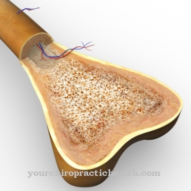
In addition to the growth in length, there is also growth in thickness. This process corresponds to ossification from the outside, i.e. perichondral ossification. During this process, osteoblasts separate from the skin of the cartilage (perichondrium). After detachment, they are deposited in the form of a ring around the model of the cartilage. This creates a so-called bone cuff. Perichondral ossification always occurs on the median shaft (diaphysis) of long tubular bones and corresponds to their appositional growth.
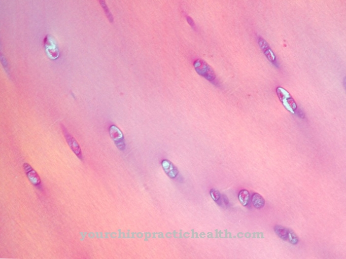
The ossification points in the context of ossification are also called ossification centers or bone nuclei. In both perichondral and enchondral ossification, the osteoblasts involved release osteoid. Osteoblastic enzymes have an influence and support the deposition of calcium salts. After these processes, the osteoblasts become osteocytes.
When bone fractures are healed, ossification processes result in braided and fibrous bones, which become more and more resilient through bone remodeling processes. During bone growth, the longitudinal growth takes place in the section of the growth plate on the middle piece, around the edge of which the perichondral bone cuffs lie.
The chondrocytes eventually multiply in the direction of the epiphysis. There is a supply of undifferentiated chondrocytes in the reserve zone. The proliferation zone contains active chondrocytes that multiply in a mitotic manner, thus forming longitudinal columns. In the hypertrophic zone, the columnar chondrocytes grow hypertrophically and mineralize the longitundinal septa.
Only in the opening zone are enzymes secreted that build the transverse septa. The longitudinal septa are ossified by osteoblasts in the opening zone. At the end of the growth phase, the dia- and epiphysis grow together.
Illnesses & ailments
Diseases related to osteogenesis are also known as bone formation disorders. This group includes, for example, mutation-related achondroplasia, which is known to be the most common cause of genetically related short stature. A point mutation in the growth factor receptor gene FGFR-3 disrupts cartilage formation. The bone growth zone ossifies prematurely and thus restricts the length growth of the arms and legs. This condition is an enchondral ossification disorder.
Most of the other bone growth disorders also mainly affect the enchondral and rather less the perichondral ossification. A second example from the same group of diseases is Fibrodysplasia ossificans progressiva, in which the connective tissue ossifies prematurely. The reason for this is a missing switch-off signal for the gene that controls skeletal growth in the development of the fetus.
In addition to endchondral ossification, glass bone disease also directly affects perichondral osteogenesis. Type I collagens are a main element of connective tissue and are correspondingly relevant for any structure of the bone matrix. In glass bone disease, a point mutation of type I collagen on chromosomes 7 and 17 changes the structure of the collagens. For this reason, the most important amino acids in collagen are exchanged for other amino acids. The collagen synthesis is reduced and the twisting of the triple helix is hindered. The collagens therefore lose their stability. The affected bones are therefore of a glassy structure and break at the slightest load.




.jpg)


.jpg)


.jpg)
