The pupil is significantly involved in the visual process. It regulates the incidence of light on the retina and is thus involved in the creation of the visual impression. Through the process of stimulus processing, it adapts to the prevailing light conditions.
What is the pupil
In the eye is that pupil visible as a black circle and forms the opening of the iris. It is a recess in the iris tissue. The pupil is also called Eye hole designated. The term is derived from the Latin word "pupilla", which means something like "doll".
The reason for this is the reduced self-reflection in the eye of the other person, which was perceived as a doll. The size of the pupil is determined by the incidence of light and its angle.
Anatomy & structure
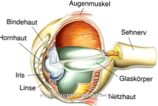
The diameter of the eye hole varies between 1.5 and 8-12 millimeters. On the outside, it is covered by the anterior chamber and the cornea. The lens is located inside the eye behind the pupil. It is controlled by the inner eye muscles: the pupil constrictor or pupil closer (Sphincter pupillae muscle) and the pupil dilator or opener (Dilator pupillae muscle).
Ring-shaped and fan-shaped muscles behind the eye are responsible for its size. The muscle contraction and the adjustment of the pupil size happens unconsciously and is dependent on the surrounding light. This adjustment is known as the pupillary reflex. A conscious control of the pupil size is not possible. It is subject to various factors.
Function & tasks
Together with the iris, the pupil functions as the diaphragm mechanism of the eye. They control the light falling on the retina. This means that the iris and pupil are involved in the first step of stimulus reception. In the eye, the light is processed as a stimulus.
The retina forwards it to the optic nerve, from where the information is transmitted to the brain. During the pupillary reflex, information is passed on to the central nervous system on the one hand (afference) and, on the other hand, the corresponding muscles are activated (efference).
Usually the pupils are the same size. This is due to the crossing of nerve fibers that lead from the midbrain to the eyes. Brightness makes the pupils smaller, darkness enlarges them. The change in brightness is perceived by the retina, but can only get used to it slowly. The pupil takes over the regulation. Medicine calls the widening of the eye hole as mydriasis, while the narrowing is also called miosis.
Both terms come from the Greek. The constriction of the pupil, also called parasympathetic innervation, is a process of the autonomic nervous system. It is sometimes responsible for the recovery and regeneration of the body. Similar to a camera, the narrowing of the pupil increases the depth of field. When narrowing, marginal rays are masked out, which avoids blurry images.
The opposite sympathetic innervation, i.e. the expansion, triggers an increase in performance of the organism. An example of this is the dilation of the pupil in the dark. This process enables the scarce light to be absorbed more effectively.
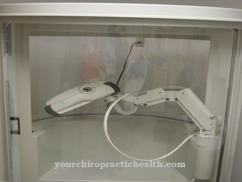
In addition to its primary function, the pupil also indicates emotions. For example, the pupils dilate with fear, disgust or joy. These aspects depend on the autonomic nervous system, which reacts to the emotional state. A new study deals with reading decisions based on the change in pupil size. In what is known as pupillometry, doctors measure this quantity using an infrared camera. This can be used to measure the person's emotional stress on the computer.
But drug consumption, medication and various illnesses also have an effect on this. Taking drugs like heroin narrows the pupil, while cannabis and LSD, for example, enlarge them. Therefore, doctors often check the pupillary reflex during physical examinations. Depending on the symptoms, a doctor will measure your diameter and your ability to react. He also checks whether both pupils react equally to stimuli and whether they are the same size.
You can find your medication here
➔ Medicines for eye infectionsDiseases
Diseases that are reflected in the pupil size are divided into afferent and efferent diseases. The term “afferent” describes the transmission of signals to the brain, while “efferent” describes the transmission from the brain to the organ.
The damage to the retina and the associated disease is afferent. Such damage leads to problems with the transmission of the collected impressions. Therefore, the pupil adjusts itself incorrectly. Reasons for this are externally inflicted injuries, diabetes or glaucoma (glaucoma). Another possibility is a detachment of the retina.
Another afferent disease is damage to the optic nerve. External influences are rarely responsible for this. Pathological changes in the cerebral vessels or pressure on the optic nerve caused by tumors can cause such damage. Inflammations such as multiple sclerosis are also possible causes. Pupillary reactions are often the result.
Efferent disorders are triggered by muscles or their nerves. For example, external injuries or Lyme disease can affect the eye muscles. The same effects can be seen in multiple sclerosis and diabetes. Pupillotonia is a disorder of the parasympathetic innervation. The mostly harmless disease triggers a different size regulation of the pupils.
Finally, Horner's syndrome also influences the pupil setting. This is a nerve damage that is triggered by the failure of the sympathetic nervous system. A unilateral miosis with a retracted eyeball or drooping eyelid is the result. Local pill disorders can also result from congenital malformations or age-related degenerative changes. A congenital malformation of the eye is the congenital absence of the iris (aniridia).


