The roentgen-Examination or X-ray diagnostics was named after its inventor, the German physicist Wilhelm Conrad Röntgen. He discovered the rays on November 8th, 1895 in the Bavarian city of Würzburg. While X-rays are also used here in Germany, these are referred to as X-rays in other countries.
History of the X-ray

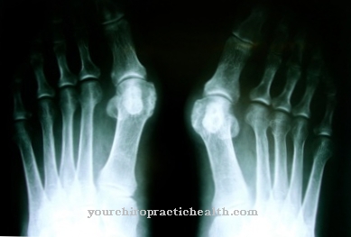
The roentgen was the first imaging technique to show bones. In the course of time, the X-ray devices have been continuously improved, so that the current dose of radiation that affects the human body during an X-ray examination is very low. Unfortunately, many medical practices still have old devices that should have been replaced a long time ago. I.
In special X-ray practices, the X-ray devices are checked every day by employees before they can be used to take the images. The latest device technology is always used here. This is currently the digital X-ray.
application
X-ray images are primarily made for colds such as bronchitis (chest x-ray) and sinusitis (x-ray of the paranasal sinuses), but also for suspected broken bones. Mammography recordings - that is, chest X-rays - are also made with special X-ray cameras. With this examination, even the smallest changes in tissue can be detected. It also shows to what extent a possible carcinoma - i.e. a benign or malignant tumor - has already spread.
There is also the option of using contrast media for X-ray examinations. This makes it possible to display organs that would otherwise not be captured by the X-rays. An examination method with X-ray contrast media is, for example, the gastrointestinal passage (MDP). Here, X-ray images are made at various intervals after the contrast agent has been taken. Through this, changes and inflammations in the gastric mucous wall, for example, but also in the small and large intestine area including the appendix, are visible.
In venography - this is an examination that can be used if a thrombosis is suspected - the X-ray examination technique is used in combination with contrast media. The contrast agent is injected into a special vein from which it is then to be transported further. If the veins, arteries or aorta become constricted somewhere, the contrast agent cannot continue to flow unhindered. This can be seen on the special X-ray images. If there is a thrombosis - i.e. a plug - an immediate emergency admission to the hospital takes place so that this blood clot can be dissolved with medication or by means of an operation.
Side effects & dangers
Every patient should have an X-ray passport with them, in which all images taken are noted. This avoids costly double examinations. Because of the X-ray examinations If only low doses of radiation are emitted, they are not individually harmful. However, if x-rays are carried out constantly, cancer can develop.
Especially with computed tomography, which also works with X-rays, you should pay attention to the entry in the X-ray passport. This has a much higher radiation dose than individual X-ray examinations.For example, a coronary CT scan, which is a computed tomography scan of the heart, has a radiation dose of around 14 millisieverts. This is 575 times the dose of a single chest X-ray examination and corresponds to around 75 percent of the annual radiation dose to which an employee in a German nuclear power plant may be exposed.
Make sure that the hospital staff comply with the X-ray guidelines. While the X-ray camera is taking pictures, no employee is allowed to be in the examination room unless he is wearing a lead apron. This is the case, for example, with contrast agent examinations. If an x-ray of the limbs or chest is done, gonadal protection should be insisted on. This prevents rays from reaching the abdomen or abdomen unintentionally.
If you take all of this into account, you don't have to worry about the radiation exposure during the next X-ray examination.




.jpg)



.jpg)


.jpg)






.jpg)
