As an independent medical discipline, the radiology Both diagnostic and therapeutic purposes through the pictorial representation of body structures. The palette ranges from classic x-rays and sonography to complex cross-sectional imaging procedures such as CT or MRI. With its various examination methods, some of which are based on contrast media, radiology offers the possibility of meaningful imaging of all body structures.
What is radiology

The radiology is a highly specialized branch of medicine that uses electromagnetic rays and mechanical waves to create images of individual parts of the body or internal organs.
The main areas of radiology, which, depending on the indication, may also use contrast media for clearer imaging, are diagnostic radiology (including its specializations such as pediatric, neurological or emergency radiology) and interventional radiology, in which therapeutic measures are carried out under radiological control.
Nuclear medicine and radiation therapy are closely related to radiology, but are regarded as independent medical sub-areas.
Treatments & therapies
Due to its variety of methods and specializations, the radiology able to offer appropriate imaging for every physical structure. Radiology is of great importance for complaints and diseases of the musculoskeletal system.
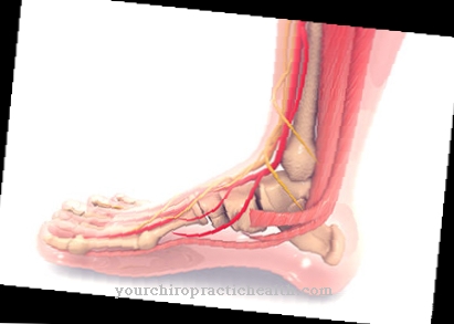
Structures such as bones, ligaments, tendons and muscles can be reliably mapped in order to subsequently initiate optimal orthopedic, surgical or physiotherapeutic treatment. The internal organs such as the gastrointestinal tract or the coronary arteries can also be reliably displayed with the available radiological examination methods.
In addition to diagnostic and therapeutic purposes, radiology also has a number of examinations in its spectrum that can be used in the context of pre- and post-operative care (for example mammography screening for the early detection of breast cancer or MRI-based clarification of surgical results). Due to rapid developments, neuroradiology, which represents the structures of the central nervous system, has become an independent branch of radiology.
Their use lies, for example, in the emergency treatment of stroke patients, aftercare after brain tumor operations or the optimal planning of intervertebral disc operations.
Diagnosis & examination methods
The modern radiology uses a large number of imaging procedures, each of which can be used not only with regard to the respective medical question, but also in coordination with special patient needs (e.g. open MRI for anxious patients or native examinations for contrast medium intolerance):
Sonography is - not least because of its low number of complications and almost any repeatability - a tried and tested standard method in radiology. Ultrasound diagnostics is an extremely gentle way of assessing organs (e.g. upper abdominal or reproductive organs) and their function, which is also suitable for pregnant women. This method is limited in obese patients and in all organs that cannot or only inadequately be visualized.
With conventional X-ray (projection radiography), radiology can image body structures (e.g. bones or thoracic organs) with the help of X-rays, whereby contrast media are often used for better assessment of organs. Examples of this are vascular representations such as angiography or venography or fluoroscopy Gastrointestinal passage after oral contrast agent intake. A common X-ray examination in the area of cancer screening is mammography, which is often offered as part of a screening.
Computed tomography (CT), like sonography and MRT, is one of the cross-sectional imaging methods in radiology. With short examination times, it provides detailed and overlay-free representations of, for example, coronary arteries or abdominal organs and, like MRI, is often used in tumor diagnostics. Because of the high radiation exposure, the doctor must carefully weigh the benefits.
Magnetic resonance tomography or magnetic resonance imaging (MRT) is a highly complex cross-sectional imaging method in radiology which, with the help of strong magnetic fields and, if necessary, additional contrast medium (especially gadolinium or iron oxide particles), provides excellent information, especially when displaying the central nervous system or the heart in real-time MRI. The advantage over CT is that it does not use ionizing radiation or iodine-containing contrast media, as well as the better soft tissue contrast.
As an independent sub-area of radiology, interventional radiology enables minimally invasive interventions with constant image control. The focus here is, for example, on the expansion of closed vessels, the stopping of bleeding in the gastrointestinal tract or the obliteration of certain tumors.


.jpg)







-eisenmangelanmie.jpg)

.jpg)


