Under the so-called retinal dysplasia is to be understood as a pathological malformation of the human retina. In most cases it is a genetic condition. Retinal dysplasia often manifests itself in the appearance of gray lines or points in focus, in distortions of surfaces or in the form of a retinal detachment.
What is retinal dysplasia?

© Edward - stock.adobe.com
The hereditary one retinal dysplasia is based on a defective development of the retina in the embryonic stage. A distinction is made between three types, with the slightest change occurring in the form of retinal folds. In technical language it is called multifocal RD designated.
The so-called geographical RD describes the appearance of irregular areas in focus, which arise due to the incorrectly developed retina. The most severe form of retinal dysplasia shows up in total RD. The retina becomes completely detached. In the progressive form of the disease, the photoreceptor cells in the retina inexorably perish.
Usually the rods are initially affected by this atrophy. For this reason, the person affected first noticed a kind of night blindness. The progressive retinal dysplasia leads to blindness in most cases and is a so-called hereditary disease that is inherited as an autosomal recessive trait.
causes
Retinal dysplasia has many different causes. It often occurs with hereditary genetic defects. However, medication, ionizing radiation, viral infections, trauma, or a lack of vitamin A can also be responsible for abnormal differentiation of the retina. Many different retinal degenerations lead to blindness. A fundamental distinction is made between early and late forms, with the latter based on genetic defects inherited in recessive genes.
Symptoms, ailments & signs
The human retina absorbs the necessary nutrients through the choroid. If the retina is damaged or if it becomes detached due to retinal dysplasia, the nutrient supply is interrupted or no longer functions adequately. This creates the typical symptoms. The person concerned perceives lightning bolts and black, twitching points in the field of vision more and more frequently. If the retina ruptures due to retinal dysplasia, tiny blood vessels are damaged.
This causes the typical appearance of flashes of light and flickering points. These can appear over a large area and are usually in rapid motion. Often, however, the occurrence of these symptoms is harmless and, in the differential diagnosis, relates to slight vitreous opacity. Those affected then usually have difficulties with strong brightness or with reading.
Since this phenomenon can also be accompanied by retinal dysplasia, which causes serious retinal damage, a specialist should always be consulted immediately. Here it can easily be determined whether the causes are harmless or whether there is retinal dysplasia, in which the retina may have already been torn off. If this takes place in the upper eye area, an elongated shadow usually appears in the field of vision, which extends from bottom to top.
If the lower part is affected, the person concerned usually notices dark areas in front of the eyes, which fall like a curtain from top to bottom. This can also lead to shifts. Basically, retinal dysplasia is accompanied by blurred vision. The disease requires specialist treatment, otherwise there is a risk of blindness. An ophthalmoscope quickly brings clarity.
Diagnosis & course of disease
The retina is located in the back of the human eye. For this reason, the ophthalmologist has no way of recognizing dysplasia without appropriate medical aids.Usually the so-called ophthalmoscopy, the ophthalmoscope, is used. Eye drops that enlarge pupils are used for this. After a short wait, the doctor inspects the eyes using a magnifying glass.
The fundus is illuminated and changes in the retina that occur in retinal dysplasia are easy to see. If a retinal detachment has already taken place, gray-looking folds as well as holes and tears can now be seen. If there is bleeding in the vitreous humor, examining the retina becomes difficult. In this case, sonography is usually used, which then gives a clearer picture of the changes.
Complications
Retinal dysplasia is hereditary and is associated with other organ malformations that can lead to complications. First, the retinal malformation can lead to retinal detachment. If the retina is already torn, the affected person sees an elongated shadow that runs from bottom to top in the field of vision. Without treatment, retinal dysplasia leads to blindness.
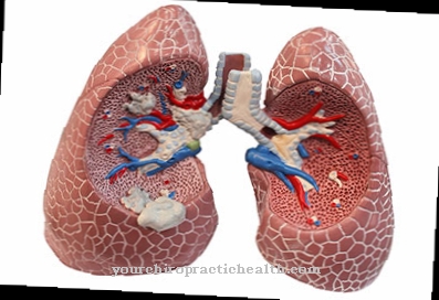
An operation is therefore often necessary to re-anchor the detached retina and at the same time to correct the changes in the vitreous humor. However, retinal dysplasia in the context of the hereditary disease is only one symptom within a whole complex of symptoms of organic malformations. In addition, there are brain and lung malformations, malformations in the gastrointestinal area, heart defects and various deformities in bones and skeleton.
The brain malformations are usually associated with a mental limitation. In severe cases it can also lead to death. The complications due to the lung malformations also depend on the type of dysplasia. In the worst case scenario, a miscarriage occurs because the lungs are not developing at all.
Otherwise severe chronic disorders of the lung function with the risk of inflammation and edema develop. Furthermore, the prognosis of the disease also depends on the type of heart defect. Overall, a patient with hereditary retinal dysplasia requires constant medical supervision to avoid complications.
When should you go to the doctor?
With this disease, a doctor must be consulted in any case, as there is no self-healing. In the worst case, the affected person can go completely blind from the disease if it is not treated in time. A doctor should be consulted if the retina of the eyes is damaged. The retina can become detached, which can lead to visual problems. The patients suffer from blurred vision or from double vision and generally from ametropia. If these symptoms appear suddenly and for no particular reason, a doctor must be consulted in any case. Visual problems in bright light can also indicate this condition and must be examined by a medical professional.
First and foremost, an ophthalmologist can be seen with retinal dysplasia. In some cases, further treatment may require surgery. Whether the disease can be treated in full cannot generally be predicted, since the further course depends heavily on the stage and the severity of the disease.
Treatment & Therapy
Treatment of retinal dysplasia is often medication. If it is diagnosed, an operation should be done as soon as possible. If the retina has already become detached, drug therapy is no longer possible. If the retina is only torn, laser treatment can be successful.
The laser beams cause inflammatory reactions, as a result of which the retinal tissue scars. This closes the damage to the retina and prevents it from becoming detached. However, if the detachment has already taken place, the laser operation is no longer successful. In this case, ophthalmic surgery is essential.
There are different approaches that depend on the type of retinal detachment due to the dysplasia and the extent to which it has already progressed. The objectives of the operation are to fix the detached retina and to correct the vitreous changes.
You can find your medication here
➔ Medicines for visual disturbances and eye complaintsprevention
Basically, retinal dysplasia can only be prevented to a limited extent. In order to prevent the serious sequelae of the disease, such as the detachment of the retina, it is imperative to see a specialist at the slightest symptoms. This has the opportunity to examine and treat the eye affected by retinal dysplasia at an early stage.
This also applies in particular to patients who already suffer from severe nearsightedness or who have cataracts. In most cases, hereditary retinal dysplasia cannot be prevented.
Aftercare
Retinal dysplasia is a congenital disease that is associated with malformations of the retina and internal organs. Follow-up care is necessary and must be initiated as early as possible. The main goals are a largely normal life for those affected and symptom relief or elimination.
Before treatment, the specialist must make a differential diagnosis, as the symptoms can also be based on other causal diseases. Therapy and aftercare depend on the organs affected. In many cases, surgery is needed to correct it.
This is where the well-known postoperative measures come into play, in which the healing is controlled. Even after a successful procedure, follow-up care continues. It is discontinued if the condition of the person concerned has remained stable even after regular checks.
In the case of inoperable damage, the focus is on relieving symptoms. For this, the patient is given medication. Follow-up care here is long-term and depends on the severity of the disease. It accompanies those affected throughout their life, as retinal dysplasia causes severe malformations.
The patient also learns how to deal with the disease appropriately on a daily basis. In addition to pain-relieving medicine, simultaneous psychotherapy is recommended if the symptoms have a negative effect on the mental health of the sick person.
You can do that yourself
Retinal dysplasia reduces the quality of vision, which is why those affected should see their ophthalmologist immediately if symptoms arise. The therapy is clarified in a detailed discussion between doctor and patient. Depending on the degree of the eye disease, the doctor may recommend drug treatment or surgery. Those affected should take the medication exactly as prescribed by a doctor.
If the retina has already peeled off, surgery is inevitable. In this case, the patients must not put up with long delays, but rather they should press for prompt treatment. You can also get more information about a possible laser treatment. Especially people who are very nearsighted or have cataracts should have regular examinations.
If the parents or other relatives suffer from the mostly genetic eye disease, the doctor should know. The earlier the disease can be diagnosed and treated. A good self-assessment is just as important as being honest with the doctor. It is natural to be afraid of the operation, but it shouldn't prevent those affected from making an appointment. Too many dangers lurk in everyday life, which can lead to accidents or mishaps in the case of visual problems.


.jpg)


