The Sarcomere is a small functional unit within the muscle: Lined up one behind the other, they form the thread-like myofibrils that are grouped together to form muscle fibers. Electrical stimulation from nerve cells causes the filaments within a sarcomere to slide into one another, causing the muscle to contract.
What is the sarcomere?
The human body has 656 muscles that perform active movements. The skeletal muscles are primarily responsible for voluntary movements, but also reacts to the reflex with the help of automated routines. These muscles are usually spindle-shaped and either attach directly to a bone or indirectly via a tendon.
Two types of muscles can be distinguished: smooth and striated. Smooth muscle tissue envelops many organs and has a surface without a clear structure. Striated muscles, on the other hand, are characterized by a striped pattern that extends across the fibers of the tissue and repeats at regular intervals.
Each of these sections is a sarcomere, which forms a contractile unit: when the muscle tenses, the fine fibers within a sarcomere slide into one another, shortening it and making the muscle as a whole contract. The longitudinal row of sarcomeres gives the myofibrils; many myofibrils form the muscle fiber with its many cell nuclei.
The muscle fibers are combined in the muscle fiber bundle and surrounded by a layer of connective tissue. It delimits the many muscle fiber bundles that make up a whole muscle from one another and enables the tissue to move flexibly and smoothly against one another. Muscles owe their sinewy appearance to this structure.
Anatomy & structure
Macroscopically, the sarcomere forms a section within the myofibril. The dark band (A band) is in the relaxed state in the middle of the sarcomere and is bounded by the light band (I band) on the right and left.
In the center is the M-line, which appears particularly dark under the microscope due to the superimposition of the sarcomere fibers. A Z-disk closes the sarcomere on both sides. The band pattern is created by the different density of the fabric within a section: In the darker areas, the thread-like filaments are pushed into one another and therefore let less light through.
The sarcomere is made up of two types of filaments: a complex of actin and tropomyosin and threads of myosin. Actin consists of spherical molecules that are closely lined up with one another, with the strand twisting slightly. A chain, to which other molecules hang occasionally, is entwined around this framework: tropomyosin. The second type of filament within a sarcomere is myosin, which in its entirety forms the dark A-band. A myosin molecule consists of two thinner chains, each with a thickening at the end, known as the myosin head. The two myosin chains wind around each other in a spiral to form a myosin filament.
Function & tasks
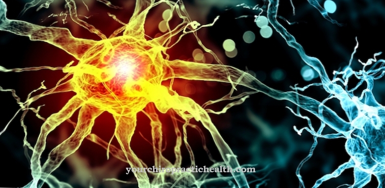
From a functional point of view, the sarcomere represents the contractile unit within the muscle. The nervous system coordinates the movement so that all the sarcomeres of a myofibril (and thus a muscle fiber) contract at the same time. A motor neuron sends an electrical signal via its nerve fiber, at the end of which there is a connection (synapse) to the muscle.
The neuron side of the synapse consists of a motor end plate in which vesicles with messenger substances (neurotransmitters) are located. The electrical signal from the nerve fiber triggers the release of neurotransmitters into the synaptic gap, on the other side of which there are postsynaptic receptors on the muscle. When a messenger substance docks to a receptor, it opens ion channels in the cell membrane through which charged particles can migrate; as a result, the electrical voltage ratio in the muscle tissue changes and an endplate potential arises.
This weak electrical current spreads through the outer membrane of the muscle cell (sarcolemma) and penetrates the interior of the tissue layer through the tube system of the T-tubules. There the electrical potential is transferred to the sarcoplasmic reticulum and allows this to release calcium ions. The calcium ions bind reversibly to the filaments of the sarcomere. The structural change allows the myosin heads to temporarily bind to the actin / tropomyosin strand and snap off.
This pushes the filament between the actin / tropomyosin threads: The sarcomere bands overlap more in this tense state than in the relaxed state, so that the sarcomere is shorter overall. The same thing happens in the adjacent sarcomeres, in many bundled muscle fibers. In larger muscles, a single motor neuron innervates several hundred muscle fibers at the same time.
You can find your medication here
➔ Medicines for muscle painDiseases
Muscle soreness is usually one of the less serious complaints that can result from slight damage to the sarcomere. Muscle soreness manifests itself in unpleasant, pulling or tearing pain in the affected muscle and a noticeable hardening of the tissue. The cause is usually due to excessive strain or insufficient warm-up during sport, which causes fine damage to the actin string.
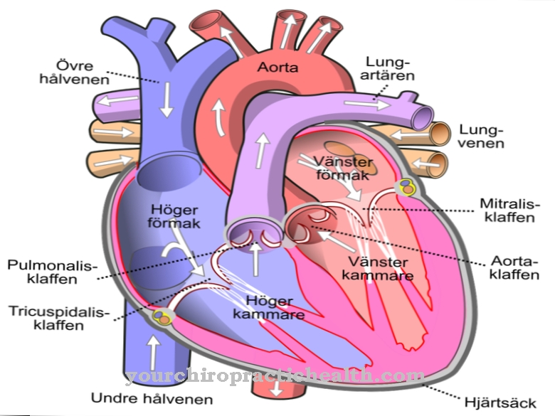
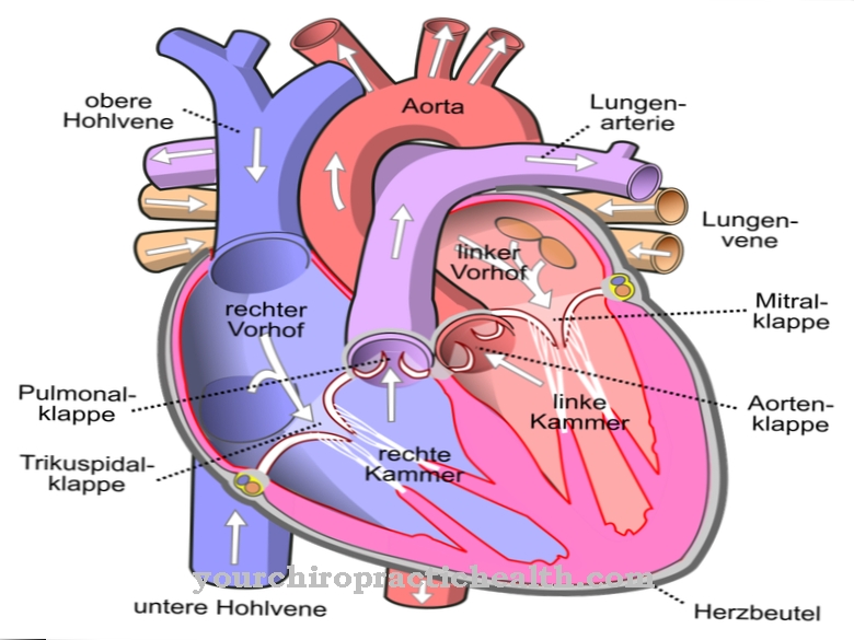
However, hypertrophic cardiomyopathy has more serious effects.In this heart disease, the sarcomeres are thicker than usual; Since fibrils and muscle fibers are still present in the same number as in a healthy person, the muscle layer is also thicker overall. This leads to functional limitations which can lead to syncope, sensation of pressure in the chest, shortness of breath, dizziness and attacks of angina pectoris. The most common causes of hypertrophic cardiomyopathy are genetic mutations, which lead to the incorrect synthesis of actin, tropomyosin or myosin in 40–60% of cases. Mutations in protein C, which binds myosin, are particularly common; this genetic defect accounts for a quarter of the causes.














.jpg)