As Cavernous sinus is an enlarged vein space within the hard meninges. He is one of the cerebral blood conductors.
What is the cavernous sinus?
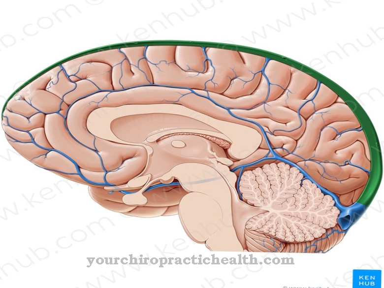
The cavernous sinus is a venous blood conductor of the human brain. The name sinus cavernosus comes from Latin.
In German, Sinus means something like “innermost”, “pocket” or “bag”. The term cavernosus is derived from the Latin word cavus (cavity or cave). The cavernous sinus is part of the cerebral blood vessels (sinus durae matris). These ensure the outflow of blood from the brain region. Various diseases can occur in the area of the cavernous sinus.
Anatomy & structure
The cavernous sinus can be found on both sides of the sella turcica (Turkish saddle), which lies on the inside of the sphenoid bone (sphenoid bone). This bone structure divides the middle cranial fossa in the area of the median plane.
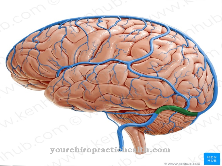
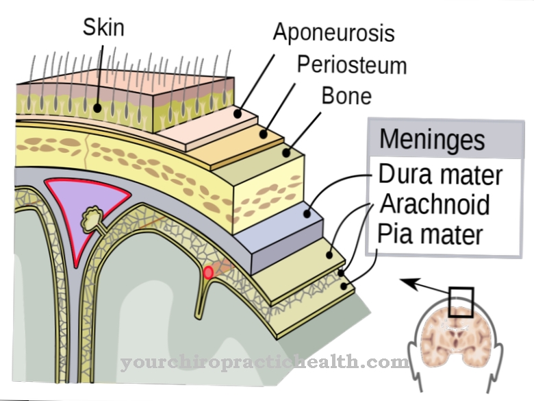
The cerebral blood conductor is located at the anterior base of the skull, where it represents a vein space within the hard meninges (dura mater). In the cavernous sinus there are inflows from the lower orbital vein (Vena ophthalmica inferior), the upper orbital vein (Vena ophthalmica superior) and the sinus sphenoparietalis. Occasionally the Sylvian vein (Vena media superficialis cerebri) is taken up by the vein space. The outflow of the cavernous sinus into the superior jugular vein takes place through the inferior petrosal sinus.
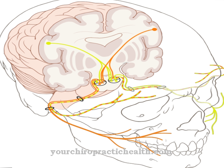
Several cranial nerves are located in the lateral wall of the enlarged venous space. These are the 3rd cranial nerve (oculomotor nerve), the 4th cranial nerve (trochlear nerve), the ophthalmic nerve (ophthalmic nerve), the maxillary nerve (maxillary nerve) and the internal carotid artery (ACI). The 6th cranial nerve, also known as the abducens nerve, runs directly through the cavernous sinus.
Function & tasks
The function of the cavernous sinus is to provide a direct passage for some important cranial nerves and the internal carotid artery, which means that they can innervate different areas of the organism. Furthermore, the cavernous sinus transports the blood from the facial area back towards the heart.
In addition, it has a role in the fact that hormones released from the adenohypophysis cross the venous space and thus get into the human body's circulation. This enables them to develop effectively. The hormones of the adenohypophysis (anterior lobe of the pituitary gland) include glandotropic and non-glandotropic hormones. While the glandotropic hormones have a stimulating effect on downstream endocrine glands, the non-glandotropic hormones have a direct effect on their target organs. Non-glandotropic hormones include prolactin and the growth hormone somatotropin (STH).
Around the cavernous sinus are the cranial nerves that control the movements of the human eyes. In addition, sensations from parts of the facial region are perceived through them.
Diseases
The cavernous sinus can be affected by several diseases and ailments. These include, for example, fractures of the skull, the formation of tumors, Tolosa-Hunt syndrome or basal meningitis.
One of the most common problems of the venous space, however, is the development of a carotid-cavernous sinus fistula. This is an abnormal connection that occurs between the cavernous sinus and a cervical (carotid) artery. The inner and outer carotid arteries supply the brain with blood. However, some people may have a tear in their arteries.If this process takes place near the cavernous sinus, there is a risk of canal formation. Doctors refer to such an unnatural canal as a fistula.
This fistula diverts the blood that normally flows through the artery into the vein. It is not uncommon for the fistula to cause increased pressure within the cavernous sinus. As a result, the affected nerves are compressed and their function is impaired. The veins leading away from the eye can also be affected by the increase in pressure. This is noticeable through visual disturbances and swollen eyes.
Doctors differentiate between a direct and an indirect carotid cavernosis sinus fistula. In a direct carotid cavernous sinus fistula, there is a connection between parts of the internal carotid artery and the veins within the cavernous sinus. This form is the most common and is characterized by an increased speed of blood flow. An indirect carotid cavernous sinus fistula is when the unnatural connection between the cavernous sinus veins and the branches within the carotid artery forms in the membranes surrounding the brain. This is noticeable by a low speed of blood flow in the fistula.
Injuries from accidents or fights as well as surgery are responsible for the development of a direct carotid-cavernous sinus fistula. In contrast, the cause of an indirect fistula is so far unknown. Another disease of the venous plexus is the cavernous sinus syndrome. The eyes of the affected person suffer from multiple symptoms of paralysis. In addition, there is a loss of sensitivity of the upper face and cornea, as well as significant headaches.
Cavernous sinus syndrome is caused by pressure damage in the cavernous sinus, which results in partial or complete failure of various cranial nerves. Possible causes are thromboses, tumors, bleeding, trauma or aneurysms on the nerve tract.
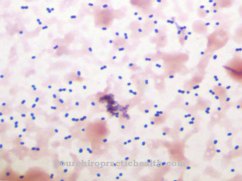
Cavernous sinus thrombosis is one of the most serious diseases of the cavernous sinus. So this can have life-threatening consequences. The thrombosis is caused by the spread of a bacterial inflammation, which in turn results from an inflammation of the frontal sinus. There is also a risk of soft tissue inflammation spreading from the upper facial region. The cavernous sinus thrombosis is noticeable through headaches, seizures, numbness in the face, chills, fever, vomiting, eye muscle paralysis and double vision.





.jpg)







.jpg)