The acoustic stethoscope is used in human medicine for listening to and auscultating various body noises. Typically, these are heart sounds, noises in the lungs and bronchi when breathing in and out, intestinal noises caused by peristalsis, and possibly flow noises in certain veins (e.g. the carotid arteries). The eavesdropping is non-invasive and the stethoscope is completely self-sufficient, i.e. independent of any electricity or other energy sources.
What is a stethoscope

The acoustic stethoscope is a non-invasive diagnostic device for making certain body noises more audible. The word stethoscope is made up of the two ancient Greek terms stethos and skopos and means something like "breast guard". A stethoscope usually consists of a head with a diameter of 30 to 46 mm, a connected tube and an ear hook that connects the two branched ends of the sound tube.
The head is used to collect the structure-borne sound like an inverted horn and forwards the sound via the sound tube to the ends of the ear hook. In the head there is usually a membrane on one side, which is set in vibrations by the incoming sound waves, similar to an eardrum, and passes them on to the air in the sound tube. There are also models in which the head can be used on both sides. As a rule, one side of the head then has a membrane and the other side has no membrane.
The membrane-free side is better suited for auscultation of deep tones, which is particularly advantageous for listening to heart sounds. The way an acoustic stethoscope works is based on simple physical-acoustic laws.
Function, effect & goals in diagnostics
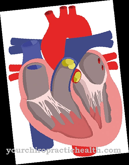
One of the main areas of application of the stethoscope is the auscultation of heart murmurs and sounds. For all four heart valves there are points near the breastbone, which, as support points for the stethoscope, allow the experienced doctor to draw conclusions about the function of the corresponding heart valve.
To the right of the sternum (to the left of the sternum from the patient's point of view) are the two points for auscultation of the mitral and aortic valve as well as the so-called Erb's point, which is suitable for acoustic diagnosis of aortic insufficiency and / or mitral valve stenosis. To the left of the sternum (from the patient's perspective, to the right of the sternum) are the two points for listening to the tricuspid valve and the aortic valve. In addition to knowledge about the quality of the function of the heart valves, auscultation of the heart sounds can also determine an atrial septal defect (ASD), a hole in the septum between the two atria and possibly existing myocarditis, an inflammation of the heart muscle.
Diagnoses made on the basis of auscultations of the heart may need to be verified by further diagnostic procedures such as EKG and ultrasound examinations. Ultrasound examinations of the heart are particularly meaningful if they are carried out esophageally, i.e. through the esophagus. The auscultated breath noises also give the experienced doctor important information about the presence of certain diseases or certain malfunctions within the respiratory system. The doctor needs a certain experience in order to be able to differentiate normal breathing sounds from abnormal or pathological breathing sounds and, above all, in order to be able to make an accurate diagnosis from perceived pathological breathing sounds.
A normal breathing sound is caused by turbulent air currents in the trachea and bronchi (central breathing sound). In addition, there are the breathing sounds, which are muffled by the lung tissue and chest wall and are often incorrectly referred to as peripheral breathing sounds. Abnormal breathing sounds can be B. be too quiet or too loud due to their development or due to impaired sound conduction, e.g. due to an accumulation of fluid (pleural effusion).
Additional breathing noises such as the typical rattle are mainly caused by liquids or secretions in the airways and require further clarification after the auscultation diagnosis. Another area of application for auscultation using a stethoscope is the two carotid arteries, the common carotid artery and the internal carotid artery, which can be affected by a pathological narrowing, a stenosis. The stenosis is usually caused by arteriosclerosis.
Especially when the stenosis - as it often happens - forms at the branching of the two carotids, the typical flow noises can be diagnosed with a stethoscope with great certainty, so that an impending stroke can possibly be averted. Auscultation of the upper abdomen can indicate a disturbed intestinal peristalsis. Typically, you should hear bowel noises about every 10 seconds. Constant loud noises or the absence of any bowel noises for several minutes indicate potentially serious disorders that must be clarified immediately with other diagnostic methods.
Risks, side effects & dangers in diagnostics
The use of the acoustic stethoscope for auscultation of certain body functions is non-invasive and free of chemical or other stresses on the body and therefore completely free of risks and side effects. A hypothetical risk can be that an inexperienced doctor makes a misdiagnosis and initiates a “wrong” therapy based on the misdiagnosis made.
During auscultation of the airways, however, it can happen that interstitial pneumonia, which initially “only” affects the supporting connective tissue between the alveoli, is not recognized because the breathing sounds are normal. In the meantime, further developed acoustic stethoscopes that work with electronic algorithms are also available. Interfering noises are muffled and the tones that are important for diagnostics are amplified.
The auscultated tones and noises can be saved on a PC and are therefore reproducible. However, these “high tech” stethoscopes seem to be gaining acceptance only very slowly, possibly because of the high price or because of (still) inadequate algorithms or because of the more complicated handling.


.jpg)


.jpg)


.jpg)


.jpg)
