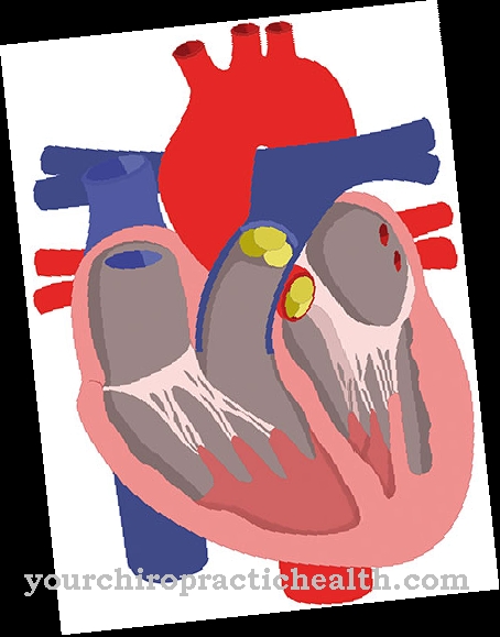
The Pulmonary artery is an artery in which oxygen-depleted blood is transported from the heart to one of the two lungs. The two pulmonary arteries are branches of the pulmonary trunk, the trunk of the lungs that connects to the right ventricle. The two pulmonary arteries are referred to as the left pulmonary artery and the right pulmonary artery for the right lung.
What is the pulmonary artery?
The pulmonary artery, too Pulmonary artery called, exists twice to conduct blood from the heart to the left and right lungs. The two pulmonary arteries represent the two branches of the branching of the pulmonary trunk (pulmonary trunk).
The two pulmonary arteries are the only arteries in which deoxygenated blood is transported.They are referred to as the left pulmonary artery for supplying the alveoli of the left lung and the dextra pulmonary artery for supplying the right lung. The pulmonary arteries open into the entrance gate (hilus) of the respective lung.
After entering the hilus, the pulmonary arteries branch further down to the level of the capillaries, which surround the alveoli, in which the exchange of substances and the oxygenation of the blood take place. The two pulmonary arteries, together with the pulmonary trunk, which forms the connection to the right ventricle of the heart, form the arterial part of the pulmonary circulation or small blood circulation.
Anatomy & structure
The two pulmonary arteries are the only two branches into which the pulmonary trunk branches. The branching (Bifurcatio trunci pulmonalis) takes place at the level of the fourth thoracic vertebra, immediately below the aortic arch. For anatomical reasons, the right pulmonary artery is somewhat longer than the left and runs below the aortic arch to the right in the direction of the pulmonary hilus, the entrance portal of the right lung.
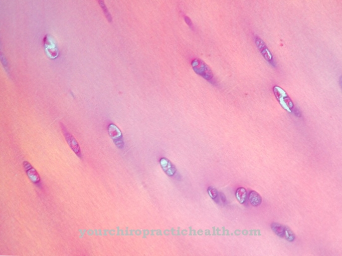
In principle, the pulmonary arteries are similar in their anatomical structure to the arteries of the body's circulation. The walls of the pulmonary artery are made up of three layers. From the inside out, these are the tunica intima, the tunica media and the tunica adventitia. The tunica intima is made up of a single-layer endothelium and an adjoining layer of loose connective tissue and the final internal elastic membrane. The tunica media is only weakly developed in the pulmonary arteries. It consists of muscle cells that wind diagonally around the blood vessels and of elastic and collagen fibers.
The tunica adventitia, which connects to the tunica media towards the outside, practically forms the supply unit for the arteries and is mainly composed of collagenous and elastic connective tissue, which is supplied with fine vessels to supply the vascular walls and with nerves to control the vasoconstriction (vasoconstriction) is streaked. However, the total vascular resistance in the pulmonary circulation is only about a tenth of the resistance of the body circulation, which has been taken into account by evolution in the anatomy of the individual layers of the vascular walls.
Function & tasks
The main task of the pulmonary arteries is to convey oxygen-poor blood from the right ventricle to the two lungs for the purpose of exchanging substances and enriching oxygen. Because the blood that is directed into the lungs is not used to supply the lungs, but to other target tissue, practically the entire body metabolism benefits, the pulmonary arteries are also known as Vasa publica.
The exchange of oxygen in the alveoli of the two lungs depends not least on the amount of oxygen in the air we breathe. If there is a lack of oxygen (hypoxia) in certain regions of the lungs, partial hypoxia triggers vasoconstriction in the arteries that are in the immediate vicinity. This means that the arterial vascular system of the lungs is individually controlled with regard to vasoconstriction. For the two pulmonary arteries, this results in a negative task, namely to react as little as possible to sympathetic impulses for arterial vasoconstriction in order not to undermine the individual cross-sectional control of the arteries within the lungs.
Diseases
In principle, functional disorders of the pulmonary arteries can be acquired or triggered by inherited genetic defects that lead to malformations of the pulmonary arteries from birth. Inherited malformations are often seen in association with other inherited heart defects.
The range of anomalies in the pulmonary arteries based on genetic defects is very large and, in rare cases, can lead to life-threatening conditions even in newborns. An acquired or inherited disease with various causes is pulmonary hypertension (PH), which occurs due to the narrowing of the blood vessels in the pulmonary arteries. In many cases, no organic cause for the occurrence of PH can be found. The mechanisms for the development and course of the disease are not yet fully understood. One of the causes is that the vascular walls react unusually strongly to messenger substances that are supposed to cause vasoconstriction, so that the vessels gradually constrict chronically and lead to the typical clinical picture.
Other causes are seen in inflammation within the vessel walls or in side effects of drugs that lead to vessel wall thickening and provoke the PH. Pulmonary embolism is another disease that can only be indirectly linked to the functioning of the pulmonary arteries. It is caused by a thrombus or embolus, a blood clot that has formed and loosened somewhere on the venous side of the body's circulation. It then enters the right ventricle with the bloodstream via the right atrium and is carried into the pulmonary circulation. Depending on its size, the thrombus then blocks one of the pulmonary arteries with potentially serious effects that can even lead to acute death.





.jpg)


.jpg)



.jpg)
