The autochthonous back muscles is the part of the back muscles that lie directly on the spine and ensure that the spine is straightened, rotated and tilted to the side, and that the head is kept upright. The term autochthonous was chosen because the musculature was created directly on the spot during the embryonic stage and not, like most skeletal muscles, “migrated” from other regions. The autochthonous back muscles are innervated by the branches of the spinal nerves on the back.
What are the autochthonous back muscles?
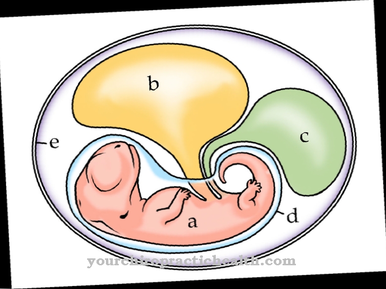
The autochthonous back muscles are already applied in place during the embryonic stage, i.e. directly adjacent to the spine and are therefore referred to as autochthonous. In contrast, many other skeletal muscles are first created in other locations before they migrate to their destination during the embryonic development phase.
The structure and function of the autochthonous back muscles do not differ from the rest of the skeletal muscles. One of the main distinguishing features of the rest of the back muscles is their innervation. The autochthonous back muscles are innervated by the branches of the spinal nerves emerging on the back (dorsal), while the remaining back muscles are supplied by the ventral (ventral) spinal nerve branches.
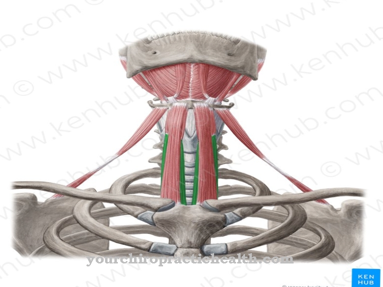
Due to the main task of the autochthonous back muscles, the individual muscles are also summarized under the term erector spinae muscle, which could be translated as "spinal erector". Overall, the autochthonous back muscles represent a very complex system of individual muscles that belong to either the lateral or the medial muscle cord (tract).
Anatomy & structure
The structure of the autochthonous back muscles does not differ from the other striated skeletal muscles that are subject to our will. The erectus spinae muscle is enveloped by the surface and deep sheets of the thoracolumbar fascia at the level of the thoracic and lumbar vertebrae and by the leaves of the nuchal fascia in the area of the cervical vertebrae.
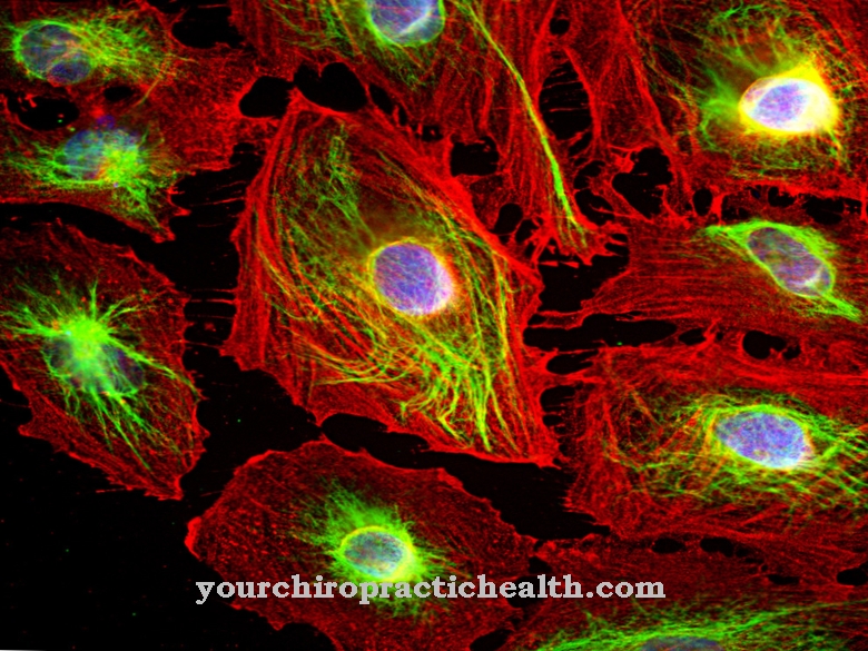
The autochthonous back muscles run in a partly bony canal, partly formed by fibers, which is formed by the bony extensions of the vertebrae or by the ribs and the enveloping fascia. The individual muscles are made up of muscle fibrils, several hundred of which each form a muscle fiber. The muscle fibers are bundled into fiber bundles, which are bundled again to form the individual muscle. The actual motor of the muscles are the myofibrils, which are composed of contractile proteins and do the actual work of contraction.
The medial muscle cord is divided into the interspinous and the transversospinal system. Interspinous muscles connect spinous processes with one another, while muscles of the transversospinal system connect the transverse processes with overlying spinous processes, whereby one or more vertebrae can also be skipped. The lateral strand of the autochthonous back muscles is divided into the intertransversal, the spinotransversal and the sacrospinal system. Usually it is about the complex muscular connection of transverse processes to one another or of spinous processes to transverse processes of different vertebral bodies.
Function & tasks
One of the main tasks of the autochthonous back muscles is to straighten the spine and head. The fanning out of the lateral and medial muscle cords into numerous individual muscles, which can inadvertently be controlled individually, allow very complex and sensitive movement sequences and patterns.
Through controlled one-sided muscle contractions of individual muscle groups, the spine can not only be bent forwards and backwards or sideways to the right or left and then straightened up again, but to a certain extent it is also possible to rotate the spine to the right and to the left. For example, the spinotransversal muscles that connect the spinous processes with the transverse processes of higher vertebrae allow the spine to be twisted in the direction of the muscle contraction in the event of unilateral contraction.
Intertransversal muscles, which connect the transverse processes with the transverse processes of the vertebrae above, allow the spine to incline in the direction of the activated muscle in the event of unilateral contraction. A bilateral contraction of the muscles leads to an extension of the spine. The deep neck muscles (muscles suboccipitales) are of particular importance. They enable fine motor movements of the head, which can be interconnected with quick messages from the sense of balance (vestibular system).
The fine motor skills of the head were originally important for humans in order to be able to better fix moving objects such as enemy or prey while moving on their own at the same time. The interaction of the various autochthonous back muscles is so complex that a certain movement of the spine is subject to voluntary control, but not the decision which muscle parts have to be brought into play by contraction or relaxation.
You can find your medication here
➔ Medicines for back painDiseases
Functional restrictions of the autochthonous back muscles are - as with other muscle parts of the skeletal muscles - either due to direct muscle diseases or to neurological problems. Diseases that only affect certain muscles of the back muscles are relatively rare.
The most common complaints are caused by muscle tension and hardening, which lead to one-sided stress on the spine and in serious cases can even trigger a herniated disc. Muscle tension in the back muscles is very common and usually causes unspecific back pain. The tension can be triggered by unusual and persistent one-sided static loads, which are intensified by permanent stress. Continuous stress or acute phases of stress that are too frequent lead to increased muscle tone due to the increased release of stress hormones, which favors muscle tension and hardening.
In rare cases, the autochthonous back muscles can also be affected by genetically caused muscular dystrophies, which lead to decreased performance of the muscles. In very rare cases, the back muscles are affected by a neuromuscular disease, in which the signal transmission from the nerve to the muscle or the sensory feedback from the muscle to the nerve is disturbed and leads to a weakening and degradation of the affected muscle.









.jpg)
