The Conjunctiva Serves as the mucous membrane layer partially on the eyeball and on the inside of the eyelids, in particular to protect the eye and the immune system. Diseases are often expressed by a reddish to brick-red discoloration of the conjunctiva.
What is the conjunctiva
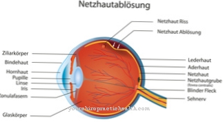
As Conjunctiva (Conjunctiva, tunica conjunctiva) is the term for the transparent, mucous membrane-like continuation of the skin in the area of the eye, which covers the lid on the rear surface to continue on the ventral (front) surface of the sclera (dermis) and then on the limbus corneae, the transition zone between the leather and the cornea to connect with the cornea.
The conjunctiva also creates a connection between the ocular bulb (eyeball) and the eyelids, with which it is firmly attached. The numerous vessels of the conjunctiva, which can be moved in a healthy state and are hardly visible to the naked eye, become more apparent when irritated due to their brick-red color.
Anatomy & structure
The Conjunctiva is usually divided into three different sections. The part of the conjunctiva that covers the back surface of the eyelid and lines its inside is called the conjunctiva palpebrarum (also conjunctiva tarsi).
This then continues as the conjunctiva fornicis with the formation of an upper and lower envelope fold (fornix conjunctivae superior or inferior) and merges into the conjunctiva bulbi, which covers the anterior surface of the sclera. At the limbus, the conjunctiva is firmly attached to the cornea. While it is firmly attached to the lids, the conjunctiva is only loosely attached to the eyeball and covers it on the ventral part up to the limbus corneae. The visible part of the sclera is completely covered by the conjunctiva.
Histologically, the conjunctiva consists of a multilayered epithelial tissue and a layer of connective tissue (lamina propria) underneath it.Within the non-keratinized epithelial layer there are also the so-called goblet cells, which are involved as mucus-forming cells in the synthesis of the tear film. The sensitive innervation of the conjunctiva is ensured primarily by branches of the trigeminal nerve.
Function & tasks

The Conjunctiva initially connects the eyeball with the lids as a transparent layer of mucous membrane (lat. "coniungere" = dt. "connect"). In addition, it acts as the outer protective covering of the eye and guarantees an additional protective mechanism through the mucus-forming goblet cells located in it, which participate in the synthesis of the tear film.
The tear film protects the eye against foreign bodies and, thanks to its antimicrobial components, the anterior globe against infections. In addition, this serves as a sliding layer for the upper lid and nourishes the vascular cornea via diffusion. The tarsal conjunctiva (conjunctiva palpebrarum) has a large number of follicle-like accumulations of plasma cells and lymphocytes, which are supposed to prevent the penetration of exogenous pathogens.
If there is inflammation, it will enlarge and form follicles that bulge (so-called follicle swelling). In addition, there are so-called Langerhans cells, especially in the tarsal conjunctiva. These cells, which are part of the dendritic system (immune defense), play an essential role in the antigen presentation through their interaction with T lymphocytes.
In addition to the corneal dendritic cells as regulators of the immune response and modulators between immune tolerance and defense, the conjunctival Langerhans cells are assigned an important function within the immune system.
Illnesses & ailments
The Conjunctiva can be affected by various impairments. One of the most common disorders is inflammatory changes in the conjunctiva (conjunctivitis), which have various causes such as chemical-physical stimuli (including foreign bodies, injuries, radiation, burns, chemical burns), bacterial (including pseudomembranous conjunctivitis, swimming pool conjunctivitis, trachomatous conjunctivitis) and viral infections (including conjunctivitis follicularis), pathological processes in the adjacent structures (including meibomian carcinoma), wetting disorders due to decreased tear secretion (including keratoconjunctivitis sicca) and allergies (including conjunctivitis vernalis) can be traced back.
Acute inflammation of the conjunctiva manifests itself symptomatically in the form of redness, swelling, heavy secretion, light sensitivity, as well as blepharospasm and chronic conjunctivitis in the form of absence of edema, reduced secretion and overgrowth of the papillary body. Since there are a large number of plasma cells, leukocytes and lymphocytes in the conjunctiva, vitreous, edematous swellings (chemosis) occur in the event of allergies, irritation, propagated inflammation (especially from the paranasal sinuses) and blood vessel congestion (e.g. due to tumors or endocrine orbitopathy) ).
After traumatic events, with severe stress (including labor, strong cough) and / or pathological changes in the blood and vascular system (including arteriosclerosis, hypotension), hyposphagmas (bleeding into the subconjunctival space) are often observed. This sub-hemorrhage is characterized by its sharp delimitation, while the conjunctiva has an intense red color. Conjunctival hemorrhage is usually harmless and is absorbed within 1 to 2 weeks.






.jpg)


.jpg)


