The Caryoplasm is the term used to describe the protoplasm within cell nuclei, which differs from the cytoplasm in particular in its electrolyte concentration. The caryoplasm creates an optimal environment for the replication and transcription of the DNA. In diabetics, cell nucleus inclusions of glycogen can be present in the caryoplasm.
What is caryoplasm?
Cell nuclei are located in the cytoplasm. They are rounded organelles of eukaryotic cells. The cell nucleus contains the genetic material of a cell. All cell nuclei are separated from the cytoplasm by a double membrane. This double matrix is called the nuclear envelope.
The genetic material is contained in it as deoxyribonucleic acid. The terms nuclear and karyo refer to the cell nuclei. The Greek term karyon means core. The caryoplasm is thus the nuclear plasma or nucleoplasm of cell nuclei. This is the entire cell nucleus content behind the nuclear envelope. The main components of the cell nucleus are chromatin, thread-like decondensed chromosomes and nucleoli. The caryoplasm is part of the protoplasm.
This refers to the cell fluid including its colloidal components. The protoplasm is made up of the caryoplasm and the cytoplasm. The living part of the cell is the cytoplasm that is surrounded by the cell membrane. The nuclear membrane separates the two forms of plasma. The main difference between the caryoplasm and the cytoplasm is the concentration of dissolved electrolytes. The karyolymph corresponds to unstructured karyoplasm. It is called core juice and is permeated by the protein structure of the core matrix. The caryoplasm interacts with the cytoplasm via nuclear pores.
Anatomy & structure
There is mainly water in the caryoplasm. Under the light microscope, it appears homogeneous in an uncolored preparation. Darker densities can appear in places.
These densities are the nuclear bodies or nucleoli and the granules of chromatin. Chromatin is the clumping and precipitation of fine chromosome fibrils. After staining, the chromocentres in them are recognizable as larger chunks. The chromatin density in the caryoplasm is dependent on cell activity. Chromatin always contains nucleoproteins, DNA, histone proteins and non-histone proteins. The junctures of the chromosome arms are called centromeres. Lighter chromatin regions correspond to loose chromatin.
Darker regions correspond to the more electron-dense chromatin areas in which the chromatin tends to clump. The lighter euchromatin of the caryoplasm must be distinguished from the electron-dense and darker heterochromatin. There is a smooth transition between the two areas. Longer parts of the unused DNA are clustered together in heterochromatin clumps of histone proteins. Function-relevant DNA sections, on the other hand, lie in the Euchromatin.
Function & tasks
Every cell is controlled from the nucleus. Almost all the genetic information of the cells is located in the caryoplasm of the cell nuclei. The genetic material of the caryoplasm is only visible during cell division and is otherwise in an unstructured form. All metabolic processes of a cell take place via RNA messenger molecules in the caryoplasm.
The caryoplasm also represents an ideal milieu for the processes of transcription and replication. During transcription, the genetic information of the cell nuclei is transferred to the RNA. This process takes place on one of the two strands. The DNA strand takes on the role of a template. Its base sequences are complementary to RNA. Transcription takes place in the cell nucleus with the help of the catalysis of DNA-dependent RNA polymerases. An intermediate product known as hnRNA is formed in the eukaryotic cells. Post-transcriptional modification turns this intermediate into mRNA.
The nuclear plasma creates the necessary environmental conditions for these processes. The same is true for the processes of replication, in which a copy of the DNA is made. The caryoplasm is not least of all mitotic. In its so-called working core, the mitotic interphase contains the user information in its non-condensed and bundled form as well as in the euchromatin network. As soon as mitosis has started in the cell nucleus, chromatin condensation takes place in the cell's caryoplasm. The chromatin is thus again in a multiple spiraled and highly ordered form and thus gives rise to the chromosomes.
Diseases
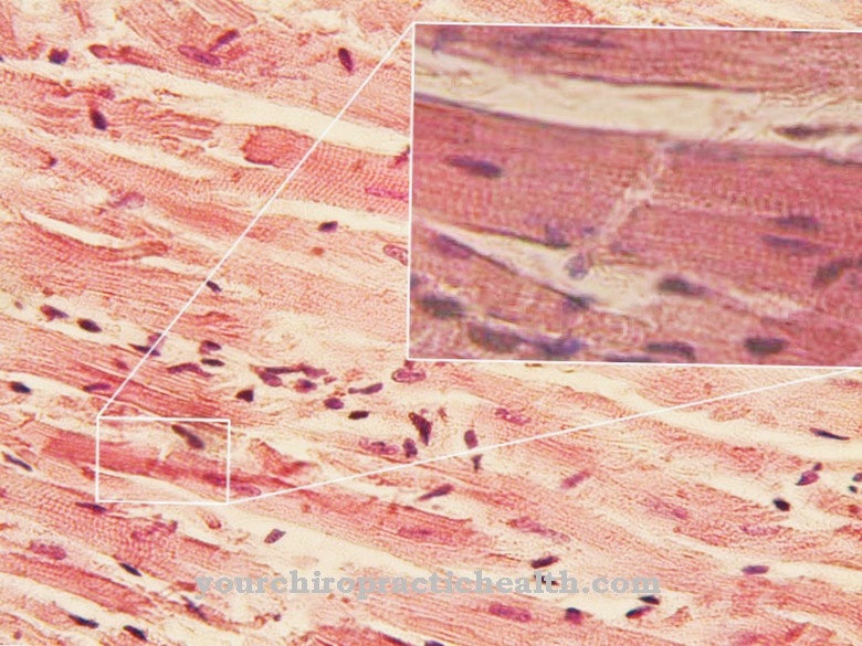
Cell damage is often examined histologically. This examination allows the type of damage to be determined more precisely. Cell damage caused by nuclear inclusions in the affected cell nuclei can often be observed in this context.
The inclusions can consist of components of the cytoplasm or foreign substances. Cytoplasmic nuclear inclusions are the most common form. They can arise from an invagination of the nuclear envelope, as can be observed in tumors. Sometimes in telophase, however, cytoplasmic structures are also included in the newly formed daughter nuclei. This phenomenon can be present in colchicine poisoning, for example. In most cases, such inclusions are separated from the caryoplasm by parts of the nuclear envelope and show degenerations. But they can also penetrate into the caryoplasm. This is often the case with glycogen deposits, as can be seen in diabetics.
Smaller particles of glycogen from the cytoplasm presumably penetrate through nuclear pores into the caryoplasm and form large aggregates there. It is possible that the caryoplasm also synthesizes the glycogen and allows it to polymerize into larger particles. In addition to infections, core inclusions are primarily associated with poisoning. The inclusions can have serious effects on mitosis. If, for example, the interphase nucleus undergoes a manifest change, negative consequences for the cells and the entire organism occur.
These relationships are discussed above all in the context of growth disorders. The caryoplasm can also completely escape from a cell nucleus when the membrane ruptures. The icing method of dermatology makes use of this connection.







.jpg)
