The Cytoskeleton consists of a dynamically changeable network of three different protein filaments in the cytoplasm of the cells.
They give the cell and organizational intracellular structures such as organelles and vesicles structure, stability and intrinsic mobility (motility). Some of the filaments protrude from the cell in order to support the motility of the cell or the directed transport of foreign bodies in the form of cilia or flagella.
What is the cytoskeleton?
The cytoskeleton of human cells consists of three different classes of protein filaments. Microfilaments (actin filaments) with a diameter of 7 to 8 nanometers, which mainly consist of actin proteins, serve to stabilize the outer cell shape and the motility of the cell as a whole, as well as intracellular structures.
In muscle cells, actin filaments enable the muscles to contract in a coordinated manner. The intermediate filaments, which are around 10 nanometers thick, also provide mechanical strength and structure for the cell. They are not involved in cell motility. Intermediate filaments consist of various proteins and dimers of the proteins, which combine to form bundles wound like ropes (tonofibrils) and are extremely tear-resistant structures. Intermediate filaments can be divided into at least 6 different types with different tasks.
The third class of filaments consists of tiny tubes, the microtubules, with an outer diameter of 25 nanometers. They are made up of polymers of tubulin dimers and are primarily responsible for all types of intracellular motility and for the motility of the cells themselves. To support the cells' own mobility, microtubules in the form of cilia or flagella can form cell processes that protrude from the cell. The network of microtubules is mostly organized from the centromere and is subject to extremely dynamic changes.
Anatomy & structure
The substance groups microfilaments, intermediate filaments (IF) and microtubules (MT), all three of which are assigned to the cytoskeleton, are almost omnipresent within the cytoplasm and also within the cell nucleus.
The basic building blocks of micro- or actin filaments in humans consist of 6 isoform actin proteins, each of which only differs by a few amino acids. The monomeric actin protein (G-actin) binds the nucleotide ATP and forms long molecular chains of actin monomers by splitting off a phosphate group, two of which connect to form helical actin filaments. The actin filaments in the smooth and striated muscles, in the heart muscles and the non-muscular actin filaments each differ slightly from one another. The build-up and breakdown of actin filaments are subject to very dynamic processes and adapt to the requirements.
Intermediate filaments are made up of various structural proteins and have a high tensile strength with a cross section of around 8 to 11 nanometers. The intermediate filaments are divided into five classes: acidic keratins, basic keratins, desmin-type, neurofilaments and lamin-type. While the keratins occur in the epithelial cells, the desmin-type filaments are found in muscle cells of the smooth and striated muscles as well as in heart muscle cells. The neurofilaments that are present in practically all nerve cells are composed of proteins such as Internexin, Nestin, NF-L, NF-M and others. Intermediate filaments of the lamin type are found in all cell nuclei within the nuclear membrane in the karyoplasm.
Function & tasks
The function and tasks of the cytoskeleton are by no means limited to the structural shape and stability of the cells. Microfilaments, which are mainly located in net-like structures directly on the plasma membrane, stabilize the outer shape of the cells. But they also form membrane protuberances like pseudopodia. Motor proteins, from which the microfilaments in the muscle cells are built, ensure the necessary contractions of the muscles.
The very high tensile strength intermediate filaments are of greatest importance for the mechanical strength of the cells. They also have a number of other functions. Keratin filaments of the epithelial cells are indirectly mechanically connected to one another via desmosomes, so that the skin tissue receives a two-dimensional, matrix-like strength. The IFs are connected to the other groups of substances in the cytoskeleton via intermediate filament-associated proteins (IFAPs), ensure a certain exchange of information and the mechanical strength of the corresponding tissue. This creates ordered structures within the cytoskeleton. Enzymes such as kinases and phosphatases ensure that the networks are built up, restructured and broken down quickly.
Different types of neurofilaments stabilize nerve tissue. Lamines control the breakdown of the cell membrane during cell division and its subsequent reconstruction. The microtubules are responsible for tasks such as controlling the transport of organelles and vesicles within the cell and organizing the chromosomes during mitosis. In cells in which microtubules develop microvilli, cilia, flagella or flagella, the MTs also ensure the motility of the entire cell or take over the removal of mucus or foreign bodies such. B. in the trachea and the external ear canal.
You can find your medication here
➔ Medicines against memory disorders and forgetfulnessDiseases
Disturbances in the metabolism of the cytoskeleton can result either from genetic defects or from externally supplied toxins. One of the most common hereditary diseases associated with a disruption in the synthesis of a membrane protein for muscles is Duchenne muscular dystrophy.
A genetic defect prevents the formation of dystrophin, a structural protein that is required in the muscle fibers of the striated skeletal muscles. The disease occurs in early childhood with a progressive course. Mutated keratins can also have serious effects. Ichthyosis, the so-called fish dander disease, leads to hyperkeratosis, an imbalance between the production and exfoliation of the skin flakes, due to one or more genetic defects on chromosome 12. Ichthyosis is the most common hereditary disease of the skin and requires intensive therapy, which, however, can only alleviate the symptoms.
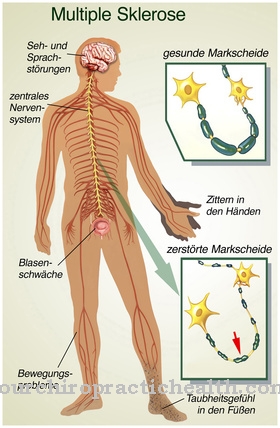
Other genetic defects that lead to a disruption of the metabolism of the neurofilaments cause z. B. amyotrophic lateral sclerosis (ALS). Some known mycotoxins (fungus toxins) such as those from mold and fly agarics disrupt the metabolism of actin filaments. Colchicine, the toxin of the autumn crocus, and taxol, which is obtained from yew trees, are used specifically for tumor therapy. They intervene in the metabolism of the microtubules.





.jpg)

.jpg)
