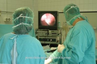
A Echocardiogram is created within echocardiography and is also referred to as "echo" for short. This is a special ultrasound examination of the heart. This procedure can be carried out in two different ways: on the one hand, as transthoracic echocardiography (TTE) or as transesophageal swallowing echo (TEE).
What is an echocardiogram?

The Echocardiogram represents an ultrasound examination of the heart, the sound waves having such a high frequency that a human ear is not able to perceive them.
The ultrasonic waves penetrate the connective tissue as well as the organs, skin and muscles. If the sound waves hit certain surfaces, they are refracted or reflected similar to light. The affected tissue areas throw the waves back with very different strengths.
An air-filled area throws them back almost completely and a liquid area absorbs the sound waves almost completely. The reflected waves can be electronically converted into an image so that the inside of the body is depicted. According to current knowledge, an echocardiogram has no damaging effect and is absolutely painless.
Function, effect & goal
Depending on which process is used, this is achieved Echocardiogram or echocardiography gives a wide range of results on the heart function and the general condition of the heart. The following information, for example, can be obtained through the echocardiogram:
- the exact size of the ventricles and atria
- the pump function or cardiac output, so that, for example, the extent of cardiac insufficiency can be assessed
- possible movement disorders in the heart muscle; can indicate a heart attack
- the function and shape of the heart valves
- Diameter and shape of the main artery (ascending aorta)
- certain changes in the pericardium, especially the importance and size of a pericardial effusion
- an estimate of blood pressure within the pulmonary artery
- as well as congenital malformations of the heart.
In an echocardiogram, a distinction is also made between the TTE and the TEE.
"Transthoracic Doppler Echocardiography", called TTE for short, is an application method with which the function and anatomy of the heart is made visible above the thorax (chest). The emitted sound waves are picked up again as echoes and visibly displayed on a screen.
The other method of application is known as "transesophageal Doppler echocardiography", or TEE for short. With this application of the echocardiogram, the examination takes place via the esophagus. This procedure is similar to a gastroscopy. The transducer is carefully guided into the interior of the esophagus so that the ultrasound waves have a shorter path to cover the heart with sound. In addition, this type of echocardiogram produces a much more precise and clearer image.
The procedure with an echocardiogram can be planned in about half an hour. The goals of this examination are an exact assessment of the heart chambers as well as the atria and valves and the pericardium. The heart continues to beat normally during the examination with an echocardiogram so that the doctor can record the full pumping actions. He can assess whether the heart valve leaflets are optimally positioned against one another and whether the heart chambers are emptying completely.
During the complete examination using the echocardiogram, an additional ECG is written in order to have a direct comparison on the ECG diagnosis strip. While the echocardiogram is being performed, the patient lies absolutely relaxed on the left side of the body.
Risks & dangers
In which Echocardiogram side effects or risks are generally extremely low. The standard TTE examination using ultrasound from the outside is neither dangerous nor unpleasant. The TEE (transosophageal echocardiography), on the other hand, could be somewhat uncomfortable. The transducer is inserted through the esophagus and positioned exactly behind the beating heart. This echocardiogram (TEE) often shows a gag reflex and increased salivation. These are natural reactions to the echocardiogram.
For example, TEE cannot be used if there are varicose veins (esophageal varices) in the esophagus, cancer of the esophagus (esophageal carcinoma) has been diagnosed, or there is an uncontrollable risk of bleeding. In addition, side effects of the locally administered narcotics (local anesthetics) may occur. The primary risk with such an echocardiogram is injury to the esophagus and throat and subsequent infections.












.jpg)

.jpg)
