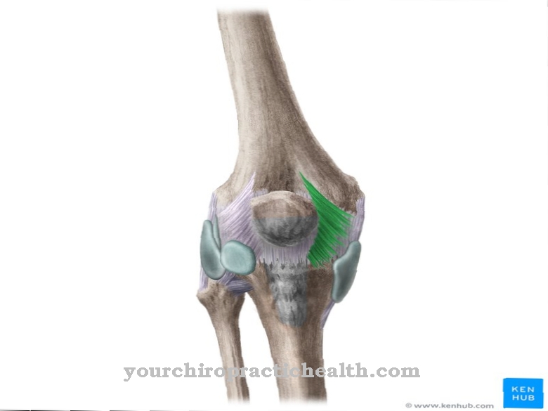
The Cubit (lat. Ulna) is a bone of the forearm that runs parallel to the spoke. Its body is diamond-shaped and consists of two end pieces, the rigid end piece forming a large part of the elbow joint and the smaller one being connected to the wrist.
What does the cubit mark?
Overall, the forearm consists of two bones: Cubit and spoke. Both are connected to one another with the help of fiber strands. The ulna lies on the little finger side and is weaker than the spoke. It consists of an elbow shaft or elbow body, the elbow head and the proximal or distal end piece.
Anatomy & structure

The bone is very close to the skin surface and is hardly protected by fatty tissue. Therefore, the bursa is located here as protection against external overloads or influences. At the front edge of the bone spur there is a depression that serves as the attachment of the joint capsule.
The process of the elbow protrudes forward like a hook. If the elbow joint is stretched, it reaches into the fossa of the bone spur, which is located on the humerus. The elbow head lies on the medial edge of the bone spur, and the elbow muscle is found on the lateral edge.
The front of the bone spur is smooth and covered by the articular cartilage, which forms part of the articular surface. The middle section of the ulna is called the ulnar shaft or the ulnar body. Together with the spoke, the ulna forms a unit because the two bones are connected to one another in different ways. On the one hand, they have an articulated connection to each other, on the other hand, a tape is stretched between them, creating an edge.
Due to its sharp edge, it can also be felt through the skin. Although the ulna is diamond-shaped, different areas can be demarcated. The front surface is the so-called bone surface, between the rear and front edge there is the surface facing the middle and the rear surface serves as the original surface for the adhesive tape. The lower end of the ulna is slightly wider and is called the ulna head.
The stylus extension is located above the wrist and the circumference of the articular surface is to the side in front. There are three variants with regard to the length ratio of ulna and radius. The most common case is that the ulna and radius are the same length. If the ulna is shorter, this is referred to as the ulnar minus, if it is longer, it is referred to as an ulnar plus.
Function & tasks
Together with the humerus, the ulna forms the elbow joint, and the stylus process also forms a small part of the wrist. The elbow joint is a hinge joint and connects the upper arm and forearm. The swivel joint between ulna and radius helps the hand and forearm to rotate.
The radius is attached to the ulna in a connective tissue ring, and the forearm rotates within this ring. The opponent to this is found in the wrist, where the ulna can turn on the spoke. In everyday life, the swivel joint is exposed to very high loads, so a ligament between the ulna and the radius - the so-called triangular fibro-cartilaginous complex (TFCC) - ensures more stability and good joint guidance. Part of that complex is that Ulnocarpal discthat stretches over the elbow head. It acts like a buffer and delimits the elbow head from the triangular leg and the moon leg.You can find your medication here
➔ Medicines for muscle painIllnesses & ailments
Cartilage damage to the head of the elbow or tears can occur, especially with heavy torsional loads or during sport Triangular disc come. The pain then mainly occurs on the little finger side of the wrist and is often aggravated when twisting with additional stress, for example when opening a lock.
Often the bursa of the elbow joint becomes inflamed. If this cannot be contained, the bursa must be surgically removed. Osteoarthritis of the elbow joint is rather rare. This occurs in patients who already have a rheumatic disease or in patients in whom the elbow joint is exposed to high physical stress.
The so-called tennis elbow is also a common disease. The tendon branch of the forearm extensor muscles is inflamed. The reason for this is usually a combination of wear and overload. Tennis players are particularly affected, but also people with very monotonous movements, such as handling tools.
Pain mainly occurs over the protruding bone when the person concerned tries to push the wrist upwards. Sometimes there is also a feeling of weakness in the wrist, which makes it difficult to grasp. In contrast to this is the golfer's elbow, where the tendon attachment of the forearm flexor muscles is inflamed.
The pain radiates here to the forearm or upper arm, and sometimes a sharp pressure pain occurs at the bone attachment. If necessary, swelling can also occur, and bending the hand or closing a fist also causes pain for the patient. The strength in the hand and finger muscles is reduced, making it very difficult to grasp. Above all, occupational groups that are exposed to constant mechanical overload are affected here.



.jpg)


.jpg)


.jpg)



.jpg)
