As External fixator a holding device is called which is used for the therapy of injured parts of the body. The treatment method is one of the osteosynthesis.
What is external fixator?

The external fixator is a holding system that is used to immobilize bone fractures. Complicated fractures associated with open wounds, in particular, are treated with this osteosynthesis procedure. The term external fixator comes from French and means “external fixation”.
An external fixator is composed of elongated screws and a rigid frame. The doctor places this outside of the body and attaches it to the affected bone with screws. The bone fragments created by the break can be stabilized in this way. In addition, they cannot shift against each other.
In the context of osteosynthesis, different procedures are used to restore broken bones. This includes the introduction of wires, screws and metal plates. However, these materials are not always suitable for open fractures because they further increase the high risk of infection. There is a risk that the germs will remain in the body, which will spread and worsen the infection. On the other hand, it makes more sense to use an external fixator with which the bone fragments can stabilize until the infection has healed.
Function, effect & goals
An external fixator is mostly used in trauma surgery to provide initial treatment for bone fractures such as debris. Typical indications are pronounced open bone fractures, a double bone fracture in the same bone, closed bone fractures in which there is severe damage to the soft tissues, and infections caused by bone fractures.
Further areas of application are polytrauma, i.e. several life-threatening injuries that are present at the same time, and pseudarthrosis. This is a so-called false joint. It forms after insufficient bone healing. Sometimes the external fixator is also used to intentionally stiffen joints. The special equipment can also be used for segment transport. The Ilisarov method, which was developed by the Soviet surgeon Gavril Ilisarov, who lengthened bones with an external ring fixator, is mostly used.
Cutting the bone at a specific point creates an artificial break. Subsequently, both bone parts are attached to an apparatus, whereby the gap at the fracture point widens increasingly. As the bone is pulled apart, it grows. Over the years this process has been improved even further.
The areas of application of the external fixator also include fractures of the cervical spine and various deformities in which it is used for callus distraction. These are mostly different leg lengths.
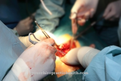
Before an external fixator is attached, the patient is given general anesthesia. How the victim is stored depends on the injury. For example, if the wrist breaks, the doctor will slightly bend the patient's arm and raise it slightly. During the procedure, the surgeon constantly checks the patient using X-rays. In this way it can be determined whether the bone fragments are also brought into the correct position by the external fixator. For this purpose it is necessary that the storage table has a permeability for X-rays. The patient's skin must be carefully disinfected. Furthermore, the patient is covered with sterile cloths.
If the bone fragments have shifted during the break, their correct position in relation to one another can be impaired. The surgeon brings them back into their correct position by pulling on them. Then some small skin incisions are made in the area of the injured bone. This gives the surgeon access to the bone. Holes are also drilled into the bone through the cuts. The surgeon then screws elongated metal rods into the holes, which connect the outer frame of the external fixator to the bone.
The appliance is attached to the bone with punch screws. They are connected to a power carrier via special jaws. The screws are inserted percutaneously. The connection force carrier is located outside the soft tissues.
After the external fixator has been attached, an X-ray examination of the patient takes place. If all bone fragments are in the desired position, the doctor can cover the inlet points of the metal rods aseptically in order to counteract infection. The patient is then taken to a recovery room where he recovers from the procedure.
Risks, side effects & dangers
Attaching an external fixator involves certain risks. This can lead to unforeseen incidents due to the anesthesia, nerve injuries and bleeding. Furthermore, the development of unsightly scars and wound infections are possible.
In addition, there is a risk of special complications. These include misalignments, infections in the bones, delays in bone healing and permanent pronounced restrictions on the movement of neighboring joints. However, if treatment is carefully planned, complications can often be counteracted.
After the operation, the patient starts physiotherapy two to three days later. In the hospital he is introduced to exercises by the physiotherapist, which he can then carry out in his own four walls. Two to six weeks later, the doctor will do additional x-rays. Consistent maintenance of the external fixator is also important. Due to the metal rods there is a risk that the wound cavity will be affected by germs. For this reason, it is necessary to carefully clean the sticks with disinfectants. In addition, the wound must remain dry.


