The Pterygopalatine fossa is an indentation in the human skull. It is located between the sphenoid bone and the upper jaw. Alternatively, it is called the palatal fossa.
What is the pterygopalatine fossa?
The pterygopalatine fossa is a part of the human skull. It is a bulge or depression in the skull bone. It can be easily felt on the face of a person with the fingers. It is located on the outside of the face just below the eye.
The pit can be felt there between the sphenoid bone and the upper jaw. The pterygopalatine fossa is also known as the alar palatal fossa due to its position and appearance. Various vessels, nerve tracts and fibers run through them. The human skull is very stable and impermeable to blood vessels and nerve tracts. In order to be able to transport the stimuli picked up from the sensory organs to the brain regions, there are bulges or channels between different tissue structures in the brain.
They are used to prevent them from being crushed or moved. The bulges serve to form ganglia, for example, or to enable an exchange between different nerve tracts. The pterygopalatine fossa is responsible for the fact that nerve pathways that can go up to the orbit, the human eye socket, can be drawn. There they then take care of the eye. In addition, it is of great importance for the care of the upper jaw.
Anatomy & structure
The pterygopalatine fossa is formed around it by various bones. The upper part of the bone is the sphenoid, a cranial bone in the shape of a butterfly. The pyramidal process of the palatine bone is located below.
This is the palatine bone. In the anterior area it is formed by the infratemporal facies of the maxilla. The pterygoid process delimits the pterygopalatine fossa to the rear. In the middle of the face is the perpendicular plate of the palatine bone. The bulge is open to the outside and can therefore be easily felt.
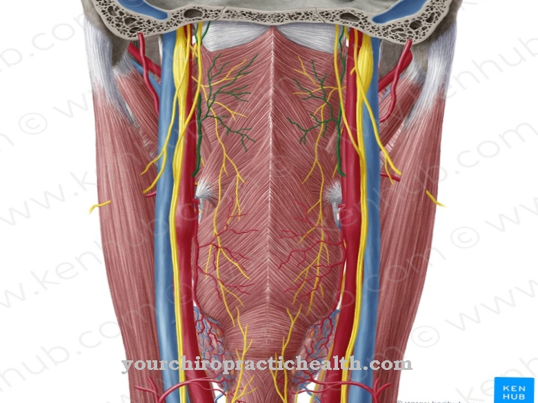
Various nerves, arteries, and veins run through the pterygopalatine fossa. They include the pterygopalatine ganglion. This is connected to the maxillary nerve. There is also the maxillary artery, also known as the pars pterygopalatina, and the zygomatic nerve. This is a terminal branch of the maxillary nerve. This in turn is a branch of the fifth cranial nerve, the trigeminal nerve. The pterygopalatine fossa also contains the major petrosal nerve and the deep petrosal nerve. Both are also known as the pterygoid canal nerve.
Function & tasks
Different stimuli such as temperature, light or touch are absorbed in the respective sensory organs and then transported to the brain via different routes. There they are evaluated and interpreted accordingly. At the same time, the various organs and brain structures are supplied via the paths used. This takes place in blood vessels with oxygen, cells or blood plasma.
Electrical signals are transported in the nerve fibers. Accordingly, a two-way exchange of information and supplies is carried out on the various communication channels. To make this possible, the nerve fibers and blood vessels need specific routes that they can use across the human head.
Since the skull is impermeable, there are various ways of access that are used. The pterygopalatine fossa is one of the bulges present. In the depression or along other anatomical spaces, the vessels and fibers can move their paths undisturbed. They are not moved or squeezed by other organs or tissues within the skull. The bulges are cavities in which there is no other cortical tissue. Therefore, they are used, for example, to be able to pick up fibers from other nerve tracts or to guarantee the transmission of existing tracts.
The zygomatic nerve, for example, picks up efferent nerve fibers from the ganglion in the pterygopalatine fossa and then moves on to the eye socket. The orbit gets its nerves from the pterygopalatine fossa. Nerve tracts of the greater petrosal nerve also run through the pterygopalatine fossa. Its nerve fibers run along the pterygoid canal. This ends in the pterygopalatine fossa. The nerve fibers in the pterygoid ganglion then take up more and move to the lacrimal gland in order to innervate it.
Diseases
Damage to the cranial bones in the face can lead to damage to the nerve fibers or vessels. If the bones around the pterygopalatine fossa are damaged, this can mean that the bulge can no longer be used as a passage.
This means that the eye socket and thus the eyes, the lacrimal gland or the upper palate are no longer adequately supplied. This can cause the skin on your face or palate to feel numb. The lacrimal gland can no longer produce sufficient tear fluid. This means that the eye is not adequately supplied. The tear fluid has an important social function, regulates inner emotional states and protects the eye. Impurities in the eye are regulated by the tear fluid. Dehydration of the eye is accompanied by pain in visual perception.
Since the skull bone is made of a very hard material, damage is usually caused by falls, accidents or as a result of surgical interventions on the face. The bone damage is often bruises or fractures that take a few weeks to regenerate. This can result in hypersensitivity of the nerves due to inflammation. Headaches and migraines are also common side effects. As soon as the bulge is closed, the blood vessels there will jam. This can lead to the formation of blood clots. This increases the risk of a stroke.











.jpg)

.jpg)
