Anastomoses are connections between anatomical structures as they occur between blood vessels, nerves, lymph vessels and hollow organs and ensure the formation of a bypass circuit if one of the connecting links is impaired.
In surgery, the doctor creates anastomoses partly artificially, whereby a distinction is made between end-to-end, side-to-side and end-to-side anastomoses depending on the respective form of this surgical intervention. One of the most common complaints associated with anastomoses is a congestion of the portal vein, which can increase blood flow to the anastomoses in this area, causing varicose veins to form in the esophagus or around the belly button.
What is the anastomosis?
The doctor describes a connection between anatomical structures as anastomosis. Such connections occur in particular between hollow organs, blood and lymph vessels, but also play a role for nerves. Blood vessels, for example, only form anastomoses with other blood vessels and lymph vessels connect exclusively with nerves.
Surgery also sometimes calls artificially produced connections anastomoses, for example the surgically restored continuity of the gastrointestinal tract after resection of individual sections of the stomach or intestine. The operative restoration of nerve connections is also associated with the artificial creation of anastomoses. As a rule, natural anastomoses are differentiated according to organ.
On the other hand, the doctor differentiates operative anastomoses according to their shape. The differentiation according to organs results in subgroups such as vascular anastomoses, intestinal anastomoses or ureteral anastomoses. The differentiation according to shape creates groups such as the end-to-end anastomosis, the end-to-side anastomosis or the side-to-side anastomosis.
Anatomy & structure
The anatomy of an anastomosis depends heavily on its tasks and thus differs according to the respective organ or the anatomical structures that are connected by them. In the lymphatic system, for example, anastomoses connect the lymphatic vessels on the same level.
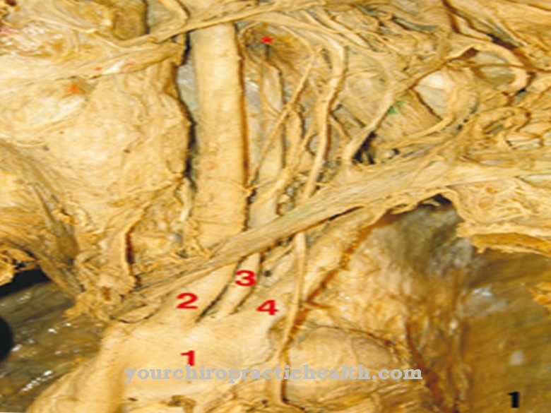
An example of an anastomosis between blood vessels, on the other hand, is the corona mortis, which is naturally abnormally developed and connects the inferior epigastric artery with the obturator artery. The Riolan anastomosis is structured differently. This inconsistent vascular connection lies in the large intestine between the arteria colica media, the arteria mesenterica superior and the arteria colica sinistra. It has an even more complex structure than the corona mortis and plays a role especially in arterial occlusions of the large intestine.
In connection with nervous anastomoses, the front teeth area of the lower jaw should be mentioned, where the nerves on the sides of the jaw are connected to each other. Artificial anastomoses can take either end-to-end, side-to-side, or end-to-side shape, meaning their anatomy is even more different. For example, in end-to-end anastomosts, the surgeon connects two sections of a hollow organ at their opened ends.
In the case of end-to-side connections, he instead sews a hollow organ section to another section that he has opened laterally. In the case of an artificial side-to-side anastomosis, two sections of a hollow organ are opened laterally in order to be sutured together.
Function & tasks
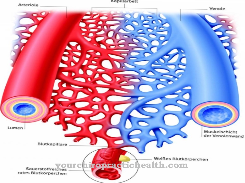
One of the most important functions of anastomoses is to create bypass circuits. This is especially true for anastomoses between vascular structures, such as the Riolan anastomosis. This connection ensures blood flow to the intestine in the event of an artery blockage in the large intestine by diverting the bloodstream from the blocked artery to another artery. In this way, anastomoses between arterial structures regulate the blood flow and, above all, prevent necrosis, which would cause tissue to die off if there was insufficient blood flow.
Anastomoses between nerves may also form bypass circuits. In this way, they secure the transmission of stimuli and thus the functional processes in the nervous system. An example of such an anastomosis is the Jacobson anastomosis. Anastomoses are also used for diversion in the lymphatic system. If the lymph flow is interrupted by vessels in one plane, for example, the anastomoses divert the lymph into an adjacent lymph vessel. In this way, the connections prevent lymphedema in the event of a flow interruption.
Diseases
High disease values can be associated with arterial anastomoses in particular. This applies above all to arteriovenous malformations, i.e. congenital malformations of the blood vessels. In the course of such malformations, arteries are sometimes directly connected to veins, which can have many threatening consequences.
Often in connection with pathological anastomoses there is also a congestion of the portal vein, in the course of which the portocaval anastomoses are supplied with more blood than usual. This can result in varicose veins in the esophagus, which are particularly risky. More rarely, this phenomenon also leads to the formation of varicose veins in the area of the navel. In addition, atypical anastomoses in the vessels of the placenta are a relatively common disease. This phenomenon is sometimes the cause of the fetofetal transfusion syndrome, which may affect identical twins.
In the case of multiple twins, atypical anastomoses in the placenta can lead to an exchange of hormones between the fetuses. If the two fetuses are of different sexes, the hormonal exchange may affect the development of the reproductive organs in the female fetus. Apart from the above, there may be many other complaints associated with anastomoses, such as fecal incontinence in an ileum-pouch-anal anastomosis. With the exception of those mentioned, almost all other anastomosis diseases are to be understood as rarities and are therefore not presented in detail.








.jpg)


.jpg)

.jpg)
