The Retinopathy prematurely (retinopathia prematurorum) is a vascular overgrowth of the retinal tissue (retina) that can occur in premature infants, especially in babies born before the 32nd week of pregnancy (SSW). Retinopathy of premature babies is divided into type 1 and type 2 and can be recognized and treated in good time by means of early detection examinations.
What is premature retinopathy?
.jpg)
© Tobilander - stock.adobe.com
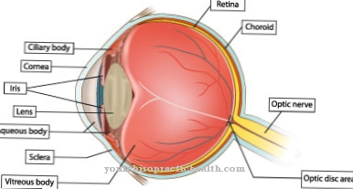
Retinopathy of premature babies is a disease of the eye. This is a damage to the retina that only occurs in premature babies. During pregnancy, the blood vessels of the retina form from the 15th week of pregnancy.
Premature birth (before the 32nd week of pregnancy) changes the oxygen supply to the blood vessels. As a result, the vessels can overgrowth, which can result in changes to the retina as well as its detachment. Depending on the type of premature retinopathy, children may need glasses or contact lenses later (often because of myopia).
However, retinopathy of premature babies can also result in more pronounced visual disturbances or even blindness. Premature babies before the 32nd week of pregnancy are particularly at risk. Babies whose birth weight is less than 1500 g or who have to be artificially ventilated for more than three days also have an increased risk of developing premature retinopathy.
causes
The cause of retinopathy in premature babies is insufficient retinal development. Since the retina and its blood vessels only start from the 15th / 16th Beginning to grow in the week, maturation is not complete until birth in the 40th week of pregnancy. Before the birth, the baby is supplied with oxygen via the mother's blood supply, so that the oxygen content in the blood is much lower than after the birth.
In premature babies, the partial pressure of oxygen increases when the child begins to breathe on its own. In the event of breathing problems, the premature baby may have to be artificially ventilated, which increases the oxygen partial pressure even further.
Because of this excess of oxygen, the sensitive, as yet immature retina is damaged, the blood vessels begin to overgrow and can sometimes even grow into the vitreous humor and cause profuse bleeding there. Another danger with retinopathy of premature babies is detachment of the retina.
Symptoms, ailments & signs
The retinopathy of premature infants caused by an increased partial pressure of oxygen during oxygen ventilation of premature infants is divided into five stages. Up to stage II it is a mild form of retinopathy, which can also regress again. However, if the change in the retina is more advanced, irreversible damage can occur, which can only be prevented through timely treatment.
In stages I and II of retinopathy of premature infants, a demarcation line or a raised border wall between the mature and immature retina develops. From stage III of the disease, new vessels and connective tissue growths form on the edge of the border wall. The newly formed vessels grow from the retina into the vitreous humor. In stage IV a partial retinal detachment takes place.
Stage V is characterized by complete retinal detachment. If left untreated, retinopathy of premature babies can lead to blindness.But later complications are also possible with treatment or with mild courses. So ametropia can develop in which distant objects can only be seen blurred (myopia).
Furthermore, the balance of the eye muscles can be disturbed with the development of a strabismus (strabismus). Glaucoma can also develop because the connective tissue growths increase intraocular pressure. In very rare cases, retinal detachment occurs years later.
Diagnosis & course
The retinopathy of premature babies is diagnosed by the ophthalmologist or pediatrician. Atropine is administered as a drop into the eyes, which causes the pupils to dilate. When the pupil has completed dilating, additional eye drops containing an anesthetic are administered.
The eye is kept open with a so-called eyelid lock. The retina of the child is examined by means of a so-called ophthalmoscopy (ophthalmoscope). The examination is usually carried out on premature babies from the 6th week of life. This examination should be checked several times.
The course of retinopathy in premature babies can be described as good. If the disease is recognized and treated in good time, it has a good prognosis. The retinopathy of premature babies type 2 usually heals completely, in exceptional cases small scars can remain on the retina, which can lead to myopia.
Type 1 retinopathy for premature infants can later lead to what is known as a secondary retinal detachment, sometimes not until years later. If not treated in time, those affected can go blind in the long run. In order to rule out long-term effects of retinopathy in premature babies, annual check-ups by an ophthalmologist are mandatory up to at least 8 years of age.
Complications
In premature infant retinopathy, also known technically as retinopathia prematurorum, the immature retina suffers damage from the premature increase in oxygen in the bloodstream. The vessels contract, which means that the retina is insufficiently supplied with nutrients and growth factors. If the constriction is not removed, the vessels can close completely.
As a result of retinopathy, there is an excessive proliferation of connective tissue outside the retina, which in some cases also releases too many growth factors. These lead to an overgrowth of vessels up into the vitreous humor of the eye and can cause retinal detachment. If the retinal detachment is not treated in time, it can lead to blindness.
Both eyes are usually affected by premature retinopathy. However, it is possible that the disease manifests itself to varying degrees in the eyes. The course of the disease also varies, but the greatest manifestation of the symptoms is always around the calculated due date. Even if the course of the disease is mild and there is no retinal detachment, the disease can have long-term consequences.
In addition to glaucoma, squinting, weak or nearsightedness can occur. In rare cases, a delayed retinal detachment with subsequent blindness can occur years later. To treat the disease, however, the administration of growth inhibitors would be necessary, which, however, would also stop the growth of the remaining organs.
When should you go to the doctor?
Premature babies are usually examined extensively by nursing and hospital staff shortly after delivery. In these routine examinations, all stages of development of the various human systems are carefully examined and dealt with, as they are not yet fully developed. As a rule, these measures are used to identify premature retinopathy at an early stage.
However, if changes are noticed in the newborn that the attending physicians have not explicitly pointed out, a discussion with the pediatrician should be sought. If the child's visual impairment is noticed or if the behavior of the premature baby is unusual, this is considered worrying.
If parents find that the child does not react to visual stimuli, they should pass this observation on. Check-ups are necessary to determine the cause. If the relatives can perceive abnormalities and in particular discoloration on the retina in the child's eye, these must be reported to the employees of the children's ward in the hospital.
Eye bleeding or unusual fluids leaking from the eye should be examined and taken care of. If there are deformations or other abnormalities of the retina or the eye, it is advisable to consult a doctor. If the retina becomes detached or cracks in the retina can be seen, these perceptions should be reported to a doctor.
Doctors & therapists in your area
Treatment & Therapy
Treatment of retinopathy prematurely depends on several factors. First, it must be determined what type of retinopathy is present in premature babies and what stage the damage to the retina is at. Type 1 is also known as premature infant retinopathy plus desease. If this “plus desease” is not present, retinopathy of premature babies is classified as type 2.
In type 2 premature retinopathy, only regular check-ups are initially carried out at very short intervals, since active therapy is not necessary here.
If type 1 retinopathy is diagnosed for premature infant, treatment must be started immediately. Depending on the severity of the retinal damage, this is treated under general anesthesia by means of cryocoagulation (icing) or laser coagulation (laser treatment).
In the case of a very severe course or secondary retinal detachment, which already leads to blindness, surgery is rarely performed nowadays.
Retinopathy of premature babies requires extensive and long follow-up care. Regular check-ups should be carried out until the disease has been successfully treated (in type 1) or until the blood vessels and retina are fully developed (in type 2 retinopathy of premature babies).
Outlook & forecast
If left untreated, retinopathy of premature babies can lead to complete blindness. If surgical measures are taken when the retinal detachment has already taken place, these show moderate success. For a good prognosis, the disease should be recognized and treated as early as possible. But even if the treatment is initially successful, long-term effects can still occur in adulthood.
Mild forms of retinopathy that have not detached the retina can resolve completely. However, many sufferers remain very nearsighted due to disease-related scars in the retina. A distortion of the retinal vessels and a displacement of the macula (point of sharpest vision) can also cause the patient to squint. Pathological, rapid eye movements (nystagmus) can also occur.
Possible long-term consequences of retinopathy of premature babies are early onset cataracts (clouding of the lens of the eye) and glaucoma (pressure damage to the eye). Scarred shrinkage of the entire eye can also lead to complete blindness on the affected side.
Due to the strain on the retina, it can develop holes or become detached years after the disease. Retinal folds can also form or other retinal changes take place. Regular checks by the ophthalmologist are recommended so that possible late effects can be recognized and treated at an early stage.
prevention
Preventive measures cannot be taken in the case of retinopathy in premature infants. In the case of artificial respiration, it is important to regularly check the oxygen content of the blood. With the help of an ophthalmological screening, however, a severe course of retinopathy of premature babies can be prevented. The screening is carried out on all premature babies born before the 32nd week of pregnancy. This means that retinopathy in premature babies can be recognized in good time and treated successfully.
Aftercare
Regular ophthalmological check-ups are necessary for mild forms of the disease that do not require treatment, as well as after therapy for retinopathy of premature babies. These take place at approximately weekly intervals until the disease has regressed significantly. However, the number and intervals of the examinations must be adapted to the individual course of the disease.
If the disease progresses satisfactorily, the close-knit examinations are usually completed when the retinal vessels have matured and the calculated due date has been reached. In addition, check-ups are carried out every few months up to at least the age of six. Objective refraction determinations (objective determination of the refractive power of the eye) and orthoptic examinations are important here (orthoptics is part of the field of strabismus medicine).
However, long-term effects and complications of retinopathy of premature babies can also occur in adolescence or adulthood. These include pseudostrabismus (apparent squint), high myopia (severe nearsightedness), scars and holes in the retina, retinal detachment, pigmentation of the retina, glaucoma (glaucoma) and cataracts (cataracts).
Without timely and adequate therapy, these long-term effects can possibly lead to blindness of the patient, which is why regular lifelong ophthalmological care is necessary. The treatment of any late-stage damage can be difficult and should therefore be done at specialized treatment centers.
You can do that yourself
Parents should carefully observe their premature baby and, if there are changes in their (visual) behavior, contact the treating ophthalmologist or the clinic as soon as possible. Older premature children who can already speak should also be watched carefully by their caregivers. It may be that the child describes a change in vision. It may just as well be that it doesn't, because, for example, a retinal detachment has happened in the amblyopic bad eye and the better eye has already taken the lead.
Since retinal detachment from premature retinopathy can also occur in later childhood, adolescence or adulthood, the warning signs of retinal detachment should be observed.
The processes in the eye due to retinopathy of premature infants can neither be prevented nor controlled. The risk of retinal detachment can be reduced by avoiding pressured breathing, heavy lifting, the risk of strong shocks and falls, as in some sports or rides at the fair.
Depending on the outcome of the surgery to reattach the retina, various options are available. If a child has a visual impairment, early intervention is advisable. This will help to strengthen the child's overall personality and identify strategies for playing and learning. Extensive information on the subject can be found in the BMAS's "Guide for People with Disabilities".



.jpg)









.jpg)
