The Inner ear As a complex structure, it is primarily used for sound perception and human orientation in the room. In many cases, hearing loss correlates with disturbances in sound perception and / or transmission in the inner ear.
What is an inner ear?

The complex structure Inner ear (Labyrinth) functions primarily as a human hearing and balance organ, which in particular ensures sound perception and spatial orientation.
The inner ear located in the petrosa ossis temporalis (pars petrosa ossis temporalis) consists of a bony labyrinth (Labyrinthus osseus), which is lined by a membranous labyrinth (Labyrinthus membranaceus) and is separated from it by a space filled with perilymph.
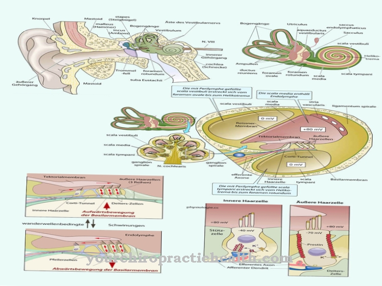
The membranous labyrinth consists of an atrium, three semicircular canals and the cochlea and is filled with so-called endolymph, a potassium-rich and liquor-like fluid. While the sensory cells in the atrium and in the semicircular canals of the inner ear serve as a vestibular organ (organ of equilibrium), the sensory cells in the snail control auditory perception.
Anatomy & structure
The hearing organ of the Inner ear is formed by the cochlea (cochlea), which is divided into three merging ducts separated from one another by membranes.
These include the membranous cochlear duct (cochlear duct) filled with endolymph, in which the organ of Corti (seat of the auditory sense) covered by the tectorial membrane is located, and that between the two other ducts, the scala vestibuli (vestibule) and the scala tympani (Timpani staircase), is located. The cochlear duct is separated by the vestibular membrane (also Reissner's membrane) from the scala vestibuli and by the basilar membrane from the scala tympani.
The vestibular organ of the inner ear, which is responsible for the sense of balance, consists of two atrial sacs, the sacculus adjoining the scala vestibuli, and the somewhat larger utriculus, located in the posterior section of the vestibulum labyrinthi (bone cavity in the temporal bone), and the posterior one the semicircular canals adjoining the vestibulum labyrinthi.
Functions & tasks
The organ of Corti inside the cochlea serves as a receptor surface made up of support cells, sensory cells and nerve fibers for sound perception, whereby the sensory cells responsible for this are also referred to as hair cells.
Sound signals arriving from outside cause a mutually opposite displacement of the basilar and tectorial membranes, so that the outer hair cells are stimulated to change their length, which amplifies the vibration of the basal membrane. As a result of the increased vibration, the inner hair cells are stimulated, which send impulses to the central nervous system via the so-called vestibulocochlear nerve (auditory nerve or 8th cranial nerve).
The vestibular organ regulates the sense of balance and is responsible for spatial orientation. The sense of rotation is regulated by the semicircular canals, which are perpendicular to one another and filled with endolymph. The rotational movement of the person in space is recorded in that the endolymph moves through the semicircular canals against the actual rotational movement of the head, whereby the hair cells there are bent. The hair cells are stimulated and send an electrical signal to the brain via the semicircular canal nerve.
The two atrial sacs that are perpendicular to each other record the translational acceleration of the person in space, with the utriculus recording the horizontal and the saccule the vertical acceleration. The information sent by the hair cells via the vestibulocochlear nerve to the brain stem is coupled and processed there with additional information coming from the eyes, spinal cord and cerebellum. In addition, the eye muscles are connected to the balance organ Inner ear interconnected, which enables a stable image with head movement.
You can find your medication here
➔ Medicines for earache and inflammationDiseases
The cochlea, whose sensitive hair cells are primarily responsible for sound perception, need sufficient nutrients and oxygen, just like the auditory nerve and the corresponding pathways in the brain.
Inadequate supply as a result of circulatory disorders can lead to corresponding functional losses. In addition, external stress (inflammation, noise, pollutants such as drugs, nicotine, alcohol or toxins) can cause irreversible damage to sound perception (sensorineural hearing loss) and in particular functional disorders of the inner ear (cochlear hearing loss).
The inner ear is often affected in old-age hearing loss (presbycusis), which is attributed, among other things, to circulatory disorders, deposits in the inner ear area as well as damaging external factors and a genetic predisposition. In addition, a disturbed sound sensation in the inner ear can cause noises in the ears such as tinnitus. Stress and stressful life situations can trigger a sudden hearing loss (acute, one-sided hearing loss).
This form of inner ear disorder can also be caused by vascular problems (insufficient blood flow and supply), infectious diseases, autoimmune reactions or benign tumors on the auditory nerve (including acoustic neuroma). In addition to other infectious diseases (meningitis, mumps, measles, herpes zoster), otitis media can spread to the inner ear without treatment and cause labyrinthitis (inflammation of the inner ear).
Menière's disease, which is still unexplained aetiologically and is characterized by the triad of symptoms occurring in the form of attacks of hearing loss, tinnitus and dizziness, is rare. Direct impairments of the vestibular organ in the inner ear also lead to balance disorders and / or dizziness.
Typical & common ear diseases
- Ear flow (otorrhea)
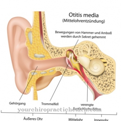
- Otitis media
- Ear canal inflammation
- Mastoiditis
- Ear furuncle










.jpg)
