Of the Carpal tunnel is a bony groove on the inside of the wrist, through which a total of 9 tendons and the median arm nerve run. The bony groove is protected from the outside by a tight band of connective tissue, the flexor retinaculum, so that a tunnel-like passage, the carpal tunnel, is formed. Common problems arise from the narrowing of the tunnel, which compresses the median nerve and causes carpal tunnel syndrome.
What is the carpal tunnel?
The carpal tunnel is formed by a special deformation of the carpal bones on the inside of the carpal joint and is limited to the outside by a tight tissue band, the retinaculum flexorum.
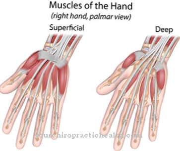
The bony groove and the tissue band, which is also known as the transverse wrist band, together form a tunnel-like passage, the carpal tunnel. It accommodates the nine tendons of the finger flexor muscles and the median nerve. The main meaning of the carpal tunnel is that the tendons of the finger flexors, even when the wrist is angled inward, are forcibly guided through the given course of the tunnel and thus run close to the body. This enormously reduces the risk of injury to the tendons if the hand is bent inwards and promotes the necessary precise fine motor skills of the fingers.
Directly below the flexor retinaculum runs the median arm nerve or nerve medianus, which contains afferent motor and efferent sensory fibers. If the tissue structures in the area of the carpal tunnel become swollen due to injuries or inflammatory reactions, the median nerve gets into a compression situation, which is the trigger for the well-known carpal tunnel syndrome.
Anatomy & structure
The bony groove of the carpal tunnel is determined by the deformation of several carpal bones. The size and shape of the gutter are largely determined by genetic predisposition. Inwardly as well as on both sides, the structure is directly adjacent to the bony membranes (periosteum) of the carpal bones.
Outwardly, the groove is covered by the flexor retinaculum, creating a tunnel-like structure. The tissue band forms a common sheath for the eight tendons of the deep and superficial flexors of the fingers and a separate sheath for the tendons of the long flexor of the thumb. In the tendon sheaths, the synovial fluid, which is also known as lubricant or synovial fluid, ensures that the tendons can move with as little friction as possible. In addition, the synovial fluid supplies the tendons and tendon sheaths with nutrients.
Above the tendons, immediately below the flexor retinaculum, the median nerve runs on the thumb side, which usually gives off a small motor branch to part of the thumb muscles within the carpal tunnel.
Function & tasks
The most important functions of the carpal tunnel are to protect and constrain the eight tendons of the finger flexor muscles and the wrist flexor on the thumb side as well as to physically protect the tendons. Without the carpal tunnel, you would have no support when the hand is flexed inward, and the conversion of the contraction of the individual finger flexors into a corresponding flexion of the fingers could not work when the hand is flexed inward.
The fact that the median nerve also runs through the carpal tunnel serves exclusively to protect the nerve mechanically, especially when the hand is flexed inwards and outwards. However, the course of the median arm nerve through the carpal tunnel directly below the flexor retinaculum is sometimes noticeable when the underlying structures “spread out” a little and put the nerve under “pressure”, meaning that there is no more space for the nerve by being displaced to let.
This can cause a typical nerve compression, which in this case is referred to as carpal tunnel syndrome. The flexor retinaculum, which delimits the carpal tunnel from the outside, is part of the hand fascia and, in conjunction with them, takes on tasks to stabilize the carpal joints and the entire wrist.
You can find your medication here
➔ Medicines for painDiseases
The most common complaints and problems observed in connection with the carpal tunnel are mostly effects of carpal tunnel syndrome.
The syndrome usually arises from inflammatory reactions on structures within the carpal tunnel. For example, tendon sheaths can become inflamed and swell slightly due to excessive strain or improper strain. This is enough to compress the median nerve and trigger typical symptoms. Because the median arm nerve carries not only motor but also sensory fibers, the first symptoms can be sensory disorders such as ants tingling in the palm of the hand or decreased sensitivity. Large parts of the palm of the hand receive sensory supply from the median nerve.
Other symptoms include motor problems and failures in the fingers and pain. For example, the index and middle fingers fail to close when trying to form a fist, a symptom known as the "oath hand". With a long-lasting carpal tunnel syndrome, an externally visible degradation of the thumb ball muscles (muscle atrophy) is also typical. The risk of developing carpal tunnel syndrome also depends on the genetically determined anatomical conditions within the carpal tunnel.
This means that the risks of developing carpal tunnel syndrome are unevenly distributed. Recurring incorrect postures such as resting the wrist on the edge of the table when using the computer mouse often lead to irritation of the median arm nerve and thus to the first symptoms of carpal tunnel syndrome. Wrist fractures or broken spokes close to the wrist are more difficult and complex. Even years later, they can narrow the carpal tunnel and cause carpal tunnel syndrome. All space-occupying changes in the wrist area such as osteoarthritis, hormonal changes, certain medications and much more can be the cause of the problems.




.jpg)


.jpg)


.jpg)
