The skeletal muscles and the visceral smooth muscles are of Motor neurons controlled that descend efferent from the CNS. The motor neurons are responsible for both reflex motor skills and all voluntary motor skills. Damage to the central motor neurons manifests itself symptomatically in so-called pyramidal trajectories.
What are motor neurons?
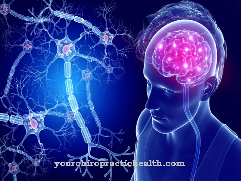
Motor neurons are motor neurons in the central nervous system. They belong to the efferent nerve cells that descend from the central nervous system. The motor neurons innervate both the skeletal muscles and the smooth muscles. The contraction of the muscles is the main task of the motor neurons. With their axons they control the muscles either directly or indirectly.
The motor neurons of the skeletal muscles are also called somatic motor neurons. They are either alpha or y neurons and are known as the lower and upper motor neurons. The a-motor neurons innervate the extrafusal muscle fibers and enable them to contract. The y-motor neurons of the skeletal muscles, on the other hand, are contained in the intrafusal muscle fibers and regulate the sensitivity of the length receptors, which transmit current information about the degree of contraction to the central nervous system.
The motor nerve cells of the smooth muscles are either specifically visceral or general visceral. In a narrower sense, only the upper and lower motor neurons of the velcro muscles are referred to as motor neurons.
Anatomy & structure
Every motor neuron receives information through the cell membrane of the dendrites and cell bodies with its receptors. This information is processed in the internal organelles and passed on chemically or electrically through the axons. For ideal conductivity, the axons are encased in a greasy insulating layer, the so-called myelin. The receptors on the cell membrane play an important role in information processing.
Transmitters in the extracellular fluid can bind to them. The receptors of the motor neurons are either ionotropic or metabotropic. After receiving information, the ionotropic receptors change the action potential at maximum speed and pass the information on rapidly. The metabotropic receptors convey information into the nucleus through numerous intermediate steps. The information is stored in the DNA in the cell nucleus. As a result, motor neurons are capable of learning processes. The synapses of the motor neurons form the transitions to the subsequent neuron.
Function & tasks
In the narrower definition, the most important task of the motor neurons is the motor control of the skeletal muscles. They are responsible for all movements of this muscular system and control both voluntary and involuntary movement sequences. Above all, the lower motor neuron in the anterior horn of the spinal cord is a superordinate control and switching point.
It primarily takes on the role of a source of inspiration. The lower motor neuron is the execution limb of all reflexes and voluntary movements that affect the skeletal muscles. With this aim, the nerve cell bodies of the lower motor neurons supply, for example, the trunk and neck muscles or the muscles of the limbs. The nerve cell bodies that supply these muscles are embedded in the gray matter of the anterior horn of the spinal cord. They stretch over the entire length of the spinal cord and form the so-called motor core column.
In the individual segments, the axons break out of the spinal canal with the help of the respective spinal nerve and thus reach the motor end plate of the respective muscles. The nerve cell bodies for the motor functions of the striated head muscles are also controlled by the lower motor neuron. However, they are not located in the spinal cord, but in the motor nuclei of the cranial nerves. The upper motor neuron is responsible for voluntary motor skills and the control of posture. The cell bodies of this motor neuron are called Betz's giant cells and are located in the motor cortex of the brain. With their axons, they shape the pyramidal trajectory and, more broadly, the extrapyramidal system.
The lower motor neuron acts as a mediator in all actions of the upper motor neuron. The voluntary motor skills are only indirectly controlled by the upper motor neuron and are closely related to the reflex motor skills.
Diseases
Motor neuron disorders impair motor skills and are often associated with an overriding loss of control over the muscles. Muscle weakness, paralysis and spasticity in particular are often the result of motor neuronal damage.
Although both spinal and cerebral infarctions can damage motor neurons, the most common causes of lesions on these nerve cell bodies are degenerative and autoimmune inflammatory diseases such as multiple sclerosis. While MS is seen as a central nervous system disease, the degenerative disease ALS explicitly affects the motor nervous system. In the disease, the motor neurons in the central nervous system break down step by step.
Lesions of the lower motor neuron, for example, paralyze the connected muscles, trigger a loss of strength or are associated with a loss of reflexes. Those of the upper motor neuron, on the other hand, are related to spastically exaggerated muscle tone in the muscles connected to it. In all motor neuronal damage, so-called pyramidal tract signs appear. These are pathological reflexes also known as the Babinski group. The reflex group corresponds to a phalanx reflex group and is interpreted to this day as providing the most meaningful indication of damage to central motor neurons.
In the infant, the reflexes of the Babinski group are not pathological, but are physiological. The signs of the pyramid orbit therefore only have a disease value from the age of around one year. Although the examination for pyramidal signs is still a standard diagnostic examination in neurology, the reliability of the pathological reflexes is now viewed critically.

