As Oculomotor nerve will the III. Called cranial nerve. He controls numerous eye movements.
What is the oculomotor nerve?
The oculomotor nerve (Ocular motor nerve) is one of the twelve paired cranial nerves. He forms the III. Cranial nerve and is responsible for the innervation of four of the six outer muscles of the eye. It also moves two inner eye muscles and the eyelid lifter.
His work is primarily done with a motor. However, there are also some parasympathetic components in it. These are noticeable during accommodation. During this process, the ciliary muscle is controlled. Together with the abducens and trochlear nerves, the oculomotor nerve also moves the eyeball.
Anatomy & structure
The oculomotor nerve has its origin in the anterior segment of the midbrain. It leaves this region of the body through the interpeduncular fossa. It crosses the dura mater (hard meninges) on the sella turcica, also known as the Turkish saddle, and runs in the ventral direction on the side wall of the cavernous sinus.
The oculomotor nerve enters the eye socket (orbit) through the upper orbital fissure (superior orbital fissure). After crossing the tin ring (annulus tendineus communis), which marks the origin of the eye muscles, the cranial nerve branches off into three branches. These are the inferior branch, the superior somatomotor branch and the ciliary ganglion, which forms a general visceromotor branch. The inferior rectus muscle supplies the inferior rectus muscle (lower eye muscle), the medialis rectus muscle (inner eye muscle) and the inferior oblique muscle (oblique lower eye muscle).
The innervation area of the superior ramus is formed by the rectus superior muscle (straight upper eye muscle) and the levator palpebrae muscle. The branch in the ciliary ganglion is connected to the postganglionic neuron. He takes care of the supply of the sphincter pupillae muscle and the ciliary muscle (ciliary muscle).
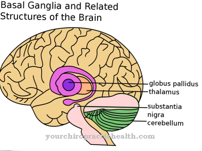
The oculomotor nerve is equipped with cranial nerve nuclei, which are called nucleus nervi oculomotorii and nucleus accessory nervi oculomotorii or nucleus Edinger-Westphal. The nucleus nervi oculomotorii forms the core of the somatomotor fibers, while this is the case with the nucleus Edinger-Westphal for the general visceromotor fibers. The somatomotor fiber core is found in the tegmentum of the midbrain (mesencephalon) at the level of the superior colliculus.
Every muscle supplied by the oculomotor nerve is equipped with its own subnucleus. However, the subnucleus of the levator palpebrae muscle is unpaired. For this reason, it is considered difficult to keep the other open when closing one eye. The nucleus acessorius nervi oculomotorii is located on the posterior side of the nucleus nervi oculomotorii.
Function & tasks
The tasks of the oculomotor nerve include supplying the eye muscles, which are important for the mobility of the eyeball. This allows the eyeball to be rotated in different directions. The muscle work is so precise that the image of the left and right eye are exactly one above the other.
Regardless of the point of view from which one sees, the same image is always fixed, which in turn guarantees spatial vision. The eye muscles and thus the oculomotor nerve are also important for accommodation, i.e. the change from near vision to far vision. In the context of accommodation, the parasympathetic part of the ocular motor nerve, which controls the ciliary muscle, becomes active. Furthermore, it narrows the iris of the pupil through the sphincter muscle. This process is called miosis.
The unpaired nucleus perlia nervus ocolumotorii is responsible for the special innervation of the ciliary muscle, which in turn enables the eye to accommodate.
You can find your medication here
➔ Medicines for eye infectionsDiseases
The oculomotor nerve can sometimes be affected by damage. One of the most common complaints is oculomotor paresis, which is a paralysis of the motor nerve of the eye. What is meant is a cranial nerve disorder that affects both men and women to the same extent.
Doctors differentiate between an external and an internal oculomotor paresis. Both unilateral and bilateral paralysis are possible. Other paralysis of the eye can also set in at the same time. The paralysis of the oculomotor nerve is caused by various causes. Most of the time, these are circulatory disorders, aneurysms or tumors within the brain stem. In some cases, oculomotor palsy is also a side effect of other diseases. These primarily include Benedict syndrome, Weber syndrome or Nothnagel syndrome.
Combination paralysis with the abducens or trochlear nerve is also possible. It is not uncommon for diabetics to suffer from paralysis of the oculomotor nerve. A significant symptom of oculomotor palsy is absolute pupillary rigidity. In addition, the patients often squint and suffer from restricted movement of the eyes or perceive double vision. Furthermore, the accommodation of the eye is restricted. If internal isolated oculomotor paralysis occurs without involvement of the external eye muscles, doctors speak of ophthalmoplegia interna.
Another typical sign of oculomotor paresis is the low level of the eye on which the paralysis occurs. There is a slight outward rotation of the eyes. Some patients also adopt a forced head position in order to maintain simple binocular vision.
A neurologist treats damage to the oculomotor nerve. The chances of recovery depend on the cause of the disease. The prognosis is considered to be more favorable if the patient suffers from circulatory disorders. In the case of aneurysms or tumors, however, an unfavorable course is to be expected. In some cases, a squint operation is performed.










.jpg)
