The medical term ossification refers to the growth of bones, also called Bone formation designated. Is a synonym ossification. It is the formation of bone tissue during the growth phase and in the callus (scar tissue to bridge the fracture gap) in secondary fracture healing in the case of bone fractures.
What is ossification?

Doctors differentiate between two types of bone formation. In desmal ossification, the bone is formed from the connective tissue; in chondral ossification, the bone is formed from an already existing cartilage.
In a normal course, ossification is a natural process of bone formation that builds up the not yet fully developed skeleton, especially in childhood. If the course is abnormal, there is increased bone formation. Bones are created where they are not intended.
Function & task
The bones develop either from the connective tissue (desmal, cranial bones, clavicles) or from the cartilage precursor (perichondral ossification). During the growth phase, bones form at the border between the metaphysis (growth in length) and the epiphysis (growth area of the long bones).
In adults, the bones renew themselves regularly through the activity of osteoblasts (bone-forming cells) and osteoclasts (bone-breaking cells).
Ossification takes place as a result of an acquired (surgery, accident, injuries) or congenital (autosomal inherited dwarfism) malformation of the skeleton due to disturbed bone formation. Overused muscles and metabolic dysregulations can also be a cause. Sometimes ossification also arises ideopathically (for no reason). It arises in the cartilaginous epiphyseal plates, which form the center of the enchondral ossification.
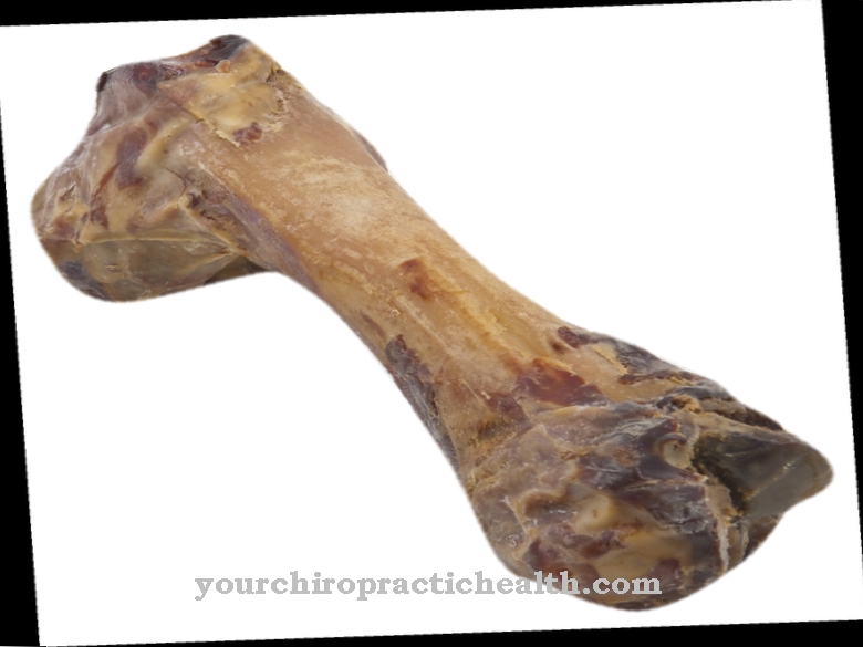
The bones of the human skeleton take various forms. There are elongated tubular bones. Your head is called the epiphysis, and the transition to the actual tubular shape is called the metaphysis. The tube as such is called the diaphysis. Typical representatives of this type of bone are the upper arm bones (humerus) and the thigh bones (femur). The skull bones are flat. The rounded sesame bones (kneecap, hand bones) form a third type of bone. The air-filled bones are the bones of the facial skull such as the sinuses.
Each bone is surrounded by a fine periosteum. Inside there is a dense bone structure (compacta, corticalis) that gives the bone strength. Uniformly oriented fibers strengthen the fabric. The bones consist of organic collagen in the form of protein, bone and fat marrow, water, phosphate and calcium.
The osteoblasts and osteoclasts in the form of small cells are located between the bone tissue. The osteoblasts are connected by fine channels and produce the bone substance. The osteoclasts act as opponents to break down the bones again. The uniformly arranged lamellar bones are responsible for the typical bone structure.
If there is a broken bone, a braided bone with fibers without any structure forms and grows haphazardly. Only with the healing process does a structured, stable lamellar bone arise again.
Desmal ossification develops from connective tissue, which is formed from mesenchymal cells. When bones grow, the cells are close together and have a good blood supply. The mesenchymal cells become osteoblasts, which produce new bones. Additional osteoblasts attach to these new, very small bones, which also form bone material, so that the bone grows appositionally through attachment. Skull bones are typically formed through this desmal process of bone growth.
In chondral ossification, bones are created as cartilage in the first step. Only in the course of this indirect (enchondral) ossification does the bone emerge from the cartilage material. Perichondral bone growth is complete from around the age of 19. The cartilage cells become larger and calcified and the osteoblasts enter the process at this point and act as building cells for bone growth.
Illnesses & ailments
Medicine distinguishes between diseases that affect regular ossification and diseases that cause excessive ossification. In patients with achondroplasia (short stature of the bones), the long bones grow in width instead of in length, as the bone growth leads to premature closure of the epiphyseal plates. The cartilage cells become larger and calcified. Since there are no more cartilage cells in the affected bone, it cannot grow in length. The vertebrae, ribs and skull bones are not affected by the achondroplasia, so that these bones develop normally, but appear larger than they are in comparison to the shortened extremities.
In heterotopic ossification, areas where connective tissue is normally found ossify. In this process, the medical term heterotopic stands for "occurring in another place". The tissue damage gives false reports to the body and causes it to produce messenger substances that lead to the ossification of cartilage tissue.
Larger bones that are affected by excessive ossification cause problems in the mechanical movement. The range of motion of the affected joints is considerably restricted. Smaller new bone formations usually do not cause any symptoms because they are too minor.
Broken bones are the most common cause of this irregular bone growth. The more complicated the fractures, the more likely it is that ossification is possible.Multiple injuries are more likely to have excessive ossification than patients with a single injury. For example, patients with hip replacement are more often affected than people with shoulder surgery. Bruising and infection can encourage excessive ossification. A fundamental prevention is not known. The orthopedic treatment starts with the axis deviations.
A lack of vitamin D in newborns has a significant impact on normal bone formation. Rickets is the most common ossification disorder in newborns. An undersupply of vitamin D automatically leads to a calcium deficiency. Since the bones consist largely of calcium, this deficiency leads to impaired bone growth. Vitamin D is therefore often given to newborns.











.jpg)
