Of the passive mass transport is the diffusion of substrates through a biomembrane. This diffusion takes place along the concentration gradient and does not require any energy. The diffusion process can be disturbed in the intestines of HIV patients, for example.
What is passive mass transport?

Cells or cell formations are separated from one another in the body by a biomembrane. Thanks to its specialized structures, this flexible separating layer enables the transport of specific molecules and information into and out of the cell interior.
There are two basic modes of transporting substances into and out of the membrane. Membranes have a selective permeability. They allow some substances to diffuse, while they act as a barrier for others.
The active transport of substances means that membranes can open up in a targeted manner for molecules to which they are actually not permeable due to their charge, concentration or size. Active transport always takes place with the use of energy. A distinction must be made between this and the passive transport type of substance transport. No energy is required for this type of movement of matter through a cell membrane. Passive transport is to be equated with diffusion processes that take place along the concentration gradient and create a concentration balance between the two sides of the membrane.
Function & task
In a cell or a cell compartment, there is a certain chemical and charge-related milieu that is necessary for the cell to function. This milieu is maintained solely by the properties of the biomembrane and the selective permeability. The passive and active substance transport supply the cell or the cell compartment in exactly the right amount with exactly the substances that are required for a beneficial environment.
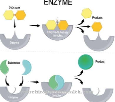
There are two different types of passive transport. The simple diffusion affects lipid soluble molecules and occurs at an extremely slow rate. They diffuse freely through the cell membrane. This form of passive transport is the one with the least effort. The second type of passive diffusion is facilitated diffusion, which in turn can be divided into two sub-forms. One of these sub-forms is carrier-mediated facilitated diffusion. With this form of passive substance transport, the membrane picks up the substrate with the help of a so-called carrier. The carrier is a protein used to identify the substance to which the substrate binds. Since simple diffusion takes place at low speed, the carrier helps transport the substance through the biomembrane. The number of all carrier molecules is limited.
For this reason, the transport through a carrier molecule is subject to saturation kinetics. The passive transport of substances by carrier molecules can also be subject to competitive inhibition. When a carrier molecule connects to its substrate, it changes its conformation and rearranges accordingly. As a result, the substrate molecule is transported through the biomembrane and only released again on the opposite side.
Some carriers can only carry one molecule at a time and thus have a uniport. Other carriers have binding sites for two different molecular substrates and only change the conformation when both binding sites are occupied. The two molecules are either symport in the same or antiport in opposite directions. There is no dependence on the electrical gradient.
The second type of facilitated diffusion is through pores and channels. This form of transport particularly affects amino acids. During ion transport, for example, the substrate of the amino acid is absorbed through pores into the cell membrane. The channels are formed by proteins. There are special binding sites on these protein-containing channels. The facilitated diffusion through pores and channels is a selective material transport that can be influenced electrically and chemically.
Almost all channels are only opened in response to certain signals. For example, a ligand-controlled channel only reacts to a messenger substance such as a hormone. Some channels are voltage controlled and open to diffusion with a change in membrane potential. After the concentration equalization, the channels close again.
Illnesses & ailments
If the membrane permeability and thus also the passive mass transport is disturbed, the permeability of various ions is no longer ideally regulated. Such membrane permeability disorders often develop from cardiovascular diseases and sometimes impair the electrolyte balance.
Sometimes membrane permeability disorders are also hereditary. Different proteins build up the biomembrane and give it a selectively permeable double lipid layer. When the proteins involved are changed, the membrane permeability also changes. This phenomenon occurs, for example, in the Myotonia congenita Thomsen. This genetic disruption of muscle function causes a gene to mutate that is responsible for coding the individual chloride channels in the muscle fiber membranes. Due to the mutation, the permeability for chloride ions is reduced and thus causes muscle stiffness.
Autoimmune diseases can also be directed against the biomembrane, such as the antiphospholipid syndrome. The immune system attacks the phospholipid-bound proteins of the membrane as part of the disease. The increased tendency for blood to clot also increases the risk of heart attacks and strokes.
Mitochondriopathies also change the permeability of the membranes. The mitochondria are the body's own energy power plants, which release free radicals when generating energy. These substances are intercepted in healthy people. This process fails in patients with mitochondiopathy, which damages the membranes and greatly reduces the ability of the mitochondria to produce energy.
The passive and active transport of substances through the membranes of the small intestine is particularly affected by disorders such as HIV enteropathy. This phenomenon particularly affects HIV patients with chronic diarrhea and can be associated with reduced activity of the interstinal enzymes.









.jpg)
