As Pleural cavity the gap between the inner and outer sheets of the pleura (pleura) is called. The pleural cavity is filled with fluid so that the two pleural leaves do not rub against each other. If there is an increase in fluid accumulation in the pleural cavity, breathing becomes difficult.
What is the pleural cavity?
The pleural cavity is called in medical terminology Cavitas pleuralis or Cavum pleurae. Since the pleural cavity is rather small, so will it Pleural space called. It lies between the wall sheet and the lung sheet of the pleura. Physiologically, there are around five to a maximum of ten milliliters of fluid within the pleural space.
Anatomy & structure
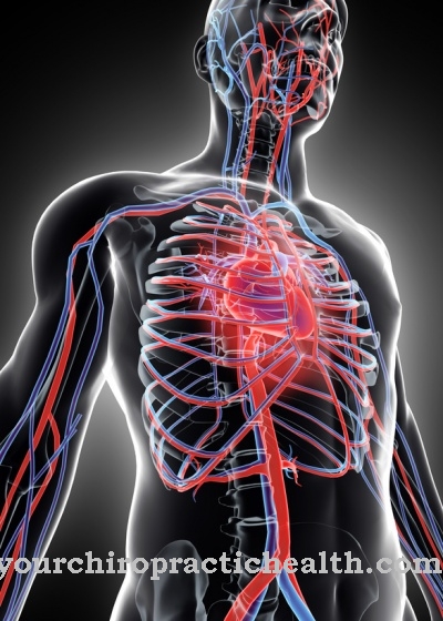
The pleura is also known as the pleura or pleura. It is a thin skin that lines the inside of the chest cavity and covers the lungs. The area that covers the lungs is called the pulmonary membrane. The pleura can be divided into four further areas.
The pleural domes lie against the lung dome. The pleura covers the inside of the ribs. The pars mediastinalis of the pleura is located in the area of the connective tissue of the middle layer and the pars diaphragmatica is located on the upper side of the diaphragm.
The pleura consists of two leaves, the visceral pleura and the parietal pleura. The visceral sheet is the inner sheet of the pleura. The parietal leaf points outwards. In the area of the lung hilus, the inner leaf merges into the outer leaf. The pulmonary hilus is the place where blood vessels, nerves, lymph vessels, and bronchi enter the lungs. The pleural cavity lies between the parietal and visceral leaves of the pleura. It is a very narrow gap rather than a cave. The gap is filled with a few milliliters of liquid. The fluid is serous, which means that it has a similar composition to the blood serum.
Function & tasks
The fluid within the pleural cavity reduces the friction between the two sheets of the pleura. The two leaves can be moved on each other, but cannot separate from each other. This can be compared to two panes of glass with a few milliliters of water between them. Due to the water film on the glass, the glass panes can be pushed back and forth on top of one another.
However, the adhesive forces prevent the two panes from being detached from one another. Since the outer sheet of the pleura adheres to the chest cavity, the inner sheet is connected to the lungs, and the two sheets in turn adhere to one another through the fluid film, the pleural space prevents the lungs from collapsing.
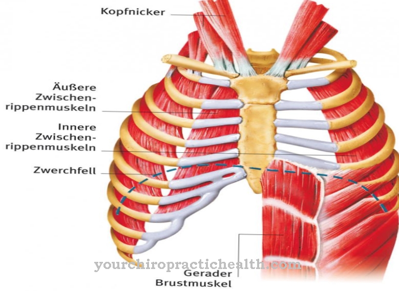
As a sliding sliding layer, the pleura with the pleural cavity is also a prerequisite for the mobility of the lungs.At the same time, it helps to create suction when you breathe in, so that the air you breathe can flow in. As the chest expands on inhalation, the outer leaf follows the pleura. The two leaves are connected by the pleural space so that the inner pleural leaf must follow the movement. Because this leaf is connected to the lungs, the lungs expand too. A negative pressure is created and the breathing air flows in.
The pressure difference between the pleural cavity and the outside air is -800 Pascal during inhalation. When exhaling, the pressure difference is reduced to -500 Pascal. If the exhalation is very forceful, the pressure within the pleura can even assume positive values for a short time.
You can find your medication here
➔ Medication for chest painDiseases
If the accumulation of fluid in the pleural cavity exceeds the physiological amount, breathing difficulties arise. Such an excessive accumulation of fluid in the pleural cavity is also known as a pleural effusion. In pleural effusions, a distinction is made between low-protein transudates and high-protein exudates.
The fluid can be bloody, purulent, or cloudy. Pleural effusions occur in the context of infectious diseases such as tuberculosis or pneumonia, can be caused by cardiac or renal insufficiency or the result of cancer. Pleural effusion can also develop after trauma or in the course of autoimmune diseases. Smaller effusions of up to half a liter of liquid are often not even noticed. The cardinal symptom with large accumulations of fluid is shortness of breath. The lungs can no longer expand properly due to the fluid in the pleural space, and as a result there is no longer enough breathing air to flow into the vessels of the lungs.
With smaller effusions, the shortness of breath only shows up during physical exertion. Larger effusions are also noticeable at rest. In addition to shortness of breath, there may be irritation of the throat or chest pain that is dependent on breathing.
If pus collects in the pleural cavity instead of fluid, it is called pleural empyema. The most common cause of pleural empyema is pleurisy, i.e. inflammation of the pleura. Haematogenic spread of pathogens and infection after trauma or after perforation of the esophagus is also conceivable. The disease is usually caused by streptococci, staphylococci, Escherichia coli, or Pseudomonas aeruginosa. Despite the accumulation of pus, the symptoms of pleural empyema may be minor. Uncharacteristic symptoms such as fever, cough and night sweats are typical.
If air gets into the pleural space, this often has life-threatening consequences. In a pneumothorax, air enters the pleural space. As a result, the two pleural leaves lose their adhesive force and the lungs collapse completely or in part. Depending on the extent of the collapse, the symptoms range from an urge to cough to life-threatening shortness of breath. The skin turns blue and there may be pain or pressure in the chest area.







.jpg)
