The Retinaculum patellae is an important part of the band system that is responsible for holding the kneecap. Its most important function is to prevent patellar luxation.
What is the patellar retinaculum?
If the translation of the Latin terms is based on the German, the term is already very aptly defined. Patella refers to the kneecap and retinaculum means the holder, accordingly we are dealing with a holder of the kneecap.
Correctly, the use of the plural is more correct, as there are a total of 3 to 4 such retaining straps on the knee. The longitudinal portions with the addition longitudinal appear regularly on the front inside and outside of the knee. The transverse reins, with the addition transversale, often exist on the outside, while they can only be detected on the inside in 30% of people. Similar straps can also be found in other parts of the human body, for example on the foot in the area of the ankle and on the upper extremity of the wrist.
Their shape and function distinguish them from the retinacula patellae. They are arranged in a semicircle and are there to attach the long tendons of the flexor and extensor muscles.
Anatomy & structure
The retinacula patellae are assigned to the connective tissue in the narrower sense. They have plenty of collagen fibers that give the structure a high tensile strength. The fibers are combined in bundles that are arranged in the main pulling direction.
The longitudinally running lateral parts arise mainly from the tendon material of the vastus lateralis muscle and the rectus femoris muscle, which are both parts of the quadriceps femoris muscle. They run close to the patella and attach laterally to the end tendon on the tibia. They are connected to the outer edge of the kneecap with connective tissue bridges.
The transverse fiber strands that pull towards the lateral surface of the thigh bone in the area of the outer ligament get their tissue material mainly from the iliotibial band, a tendon plate that extends from the pelvis over the outside of the thigh to the shin.
The inner retinacula are extensions of the tendon of the vastus medialis muscle, which also belongs to the quadriceps femoris muscle. The longitudinal reins touch the inner edge of the patella and attach to the upper edge of the tibia, medial to the quadriceps tendon. The transverse fiber strands run from the medial edge of the patella to the lateral end of the thigh bone in the area of the inner ligament.
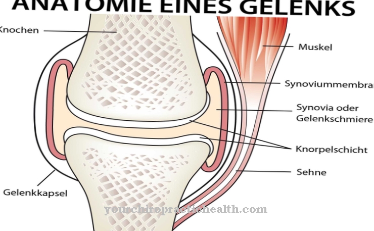
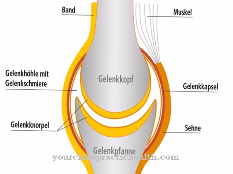
All parts are fused with the joint capsule in different areas.
Function & tasks
Together with all other tissue structures, the retinacula patellae form a thin covering layer that only insufficiently shields the underlying structures from external mechanical influences. Together with the tendons of the quadriceps, they take on a special protective function in stabilizing the knee joint. The deeper layers are fused with the capsule and strengthen it next to the kneecap and in the area of the inner and outer ligament.
All straps are very important for stability and control of the kneecap when moving. The patella runs in a groove on the front of the thigh bone. It has a matching ridge on its underside, which slides in this channel when it is bent and stretched. The bone guidance in this joint is not very pronounced, which is why other structures must take over the securing in order to prevent a dislocation of the kneecap. The retinacula patellae play a prominent role in this. The longitudinal reins that have grown together form a kind of guide rail. The transverse fibers prevent or make it more difficult for the patella to move to the opposite side. The medial parts protect against an outward dislocation, the lateral against an inward dislocation.
Since the longitudinal tracts arise from the extensor tendons and run parallel to the shin with them, they have the same function as these, but only to a lesser extent. If the patellar tendon ruptures, the quadriceps will fail completely. A small amount of residual stretching is still possible via the retinacula if they are not damaged. The term reserve stretching apparatus appears in the literature in this context.
Diseases
In certain trauma, the retinacula can also be affected. Sudden overstretching with strong flexion can tear the straps and the anterior joint capsule. The result is pain and instability of the patella with a risk of dislocation.
A comminuted fracture of the kneecap can cause the entire function of all retinacula to be lost. They lose their tension because the continuity of the bones to which they are attached no longer exists. The tightening of the joint capsule is also impaired.
A typical disease that primarily affects the kneecap, but is secondarily favored by inadequate holding ligaments, is chondropathia patellae. Often an incongruity of the two articular surfaces on the patella and the femur is the reason that the patella tends to slip outwards. If the securing ligaments and muscles are unable to prevent the displacement, dislocation can occur. The insufficiency of the ligament structures can often be traced back to a congenital weakness of the connective tissue or the consequence of a traumatic dislocation, which can lead to massive tears.
A typical sports injury, which in rare cases also affects the retinacula, which attaches to the shin, is a patellar tendon tear. This damage can be caused on the one hand by an abrupt and massive tension of the quadriceps with simultaneous flexion of the knee, as happens when suddenly stopping at full speed or when landing after a jump. On the other hand, an additional weight load during the explosive knee extension can also be responsible for the crack, as in a full span or volleyball in football. If the force is very strong, one or both of the retinacula sometimes tear off.


.jpg)


