Under the Visual pathway special somatosensitive fibers are understood that run from the retina of the eye to the visual cortex of the brain. The complex structure of the visual pathway enables human vision.
What is the visual pathway?
The visual pathway is a part of the brain. So all components have their origin in this body region. This also includes the optic nerve (nervus opticus), which is also part of the visual pathway. The neural interconnection of the optical system takes place via the visual pathway.
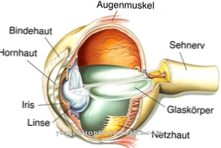
In doing so, specially somatosensitive fibers are directed from the retina of the eye towards the brain. The first member of the visual pathway is formed by the photoreceptor cells of the retina, which receive the incoming light stimuli. The cell bodies of the photoreceptor cells are located in the outer grain of the eye. They are considered the first neuron (nerve cell). From there, the nerve impulses go via the second neuron in the inner layer of the eye in the direction of multipolar retinal nerve cells within the stratum ganglionare.
The third level of nerve connections is established by these ganglion cells. With their long appendages, they form the optic nerve. The first switching of the incoming nerve impulses takes place within the retina.
Anatomy & structure
The human visual pathway has a complex structure. It extends from the posterior poles of the eyes to the cortex of the cerebrum. The retinal ganglion cells, which unite to form the optic nerve, exit in the orbit (eye socket). The optic nerve is then made up of two different fiber bundle parts.
In the right eye, the outer (lateral) part of the retina is on the right side, while the nasal part is on the left. In the left eye it is exactly the opposite. The fiber bundles of the retinal nerve cells of the respective eye attach to each other and cross each other. A little later it comes to their union in a different combination. The branching point is called optic chiasm. This is where the fibers of the nasal retinal sections cross.
Following the intersection, the fibers of the corresponding retinal sides run within the optic tract. While the right optic tract guides the fibers of the right halves of the retina, this is the case for the left optic tract with the left halves. The crossed fibers of the right eye and the uncrossed fibers of the left eye form a union in the left optic tract. This corresponds to the right half of the face. The crossed fibers of the left eye and the uncrossed fibers of the right eye, on the other hand, form their union within the right optic tract, which corresponds to the left half of the face.
Through the retinal sections, the human visual fields are reflected in opposing directions. This means that the right part of the visual field of the eyes is recorded on the left side of the retina. In contrast, the right parts of the retina represent the left halves of the visual field.
Switching between the right and left optic tracts takes place in the midbrain. From there, the so-called visual radiation goes in the direction of the cerebral cortex. Its end is located within the meningeal lobe in the center of vision on both inner sides of the brain hemispheres.
Function & tasks
The visual pathway fulfills the function of transmitting visual impressions and signals from the eye to the brain. In this way the perception of sensory impressions is made possible. Without the transmission of electrical signals to the cerebrum, humans would not be able to register the impressions seen.
Furthermore, there is a coupling between the visual pathway and the sense of balance, as well as the adjusting reflexes. If an eye impression deviates from the organ of equilibrium, the adjusting reflexes compensate for it. For example, if a person stands on a rocking ship, the fluctuations are perceived by the eyes and the organ of equilibrium. By activating the corresponding muscles, the person can continue to stand firmly. The visual pathway is divided into three functional systems. These are color and shape vision (parvocellular system), movement vision (magnocellular system) and optomotor (coniocellular system).
You can find your medication here
➔ Medicines for visual disturbances and eye complaintsDiseases
The visual pathway can be affected by various injuries or diseases. This usually creates too much pressure on the visual pathway or there is an inadequate blood supply.
Possible reasons for this are bleeding, degenerative processes, injuries, inflammation, tumors, insufficient blood flow or interruption of blood flow. Another possible cause are aneurysms, which have a bulging or widening of an artery.
Damage to the visual pathway can cause visual field defects in the affected person, which depends on which area of the visual pathway is affected. If there is a lesion of the optic nerve that leads to its disruption, this causes unilateral blindness. Doctors then speak of an amaurosis. The most common causes of this damage are optic nerve inflammation or papillary disease.
A bilateral half visual field loss on the outer side of the face can be seen in a chiasm syndrome, which is also known as the blinker phenomenon. It is mostly caused by tumors that put pressure on the optic nerve junction. Other possible causes are syphilis or multiple sclerosis. With a quick operation it is possible to regress the visual field deficits. Otherwise there is a risk of further visual disturbances.
A lateral compression of the chiasm, which is called heteronymous binasal hemianopia by medical professionals, results in hemi-lateral blindness on the same side. The reason for this is damage to the uncrossed nerve fibers. Usually sclerosis of the internal carotid artery or a bilateral aneurysm are responsible. In the case of ophthalmic migraines, shimmering scotomas are possible, which can be accompanied by headaches, dizziness, flashes of light, nausea and vomiting. In some cases, those affected also suffer from paralysis of the eye muscles. The reason for this are temporary circulatory disorders.





.jpg)


.jpg)



.jpg)
