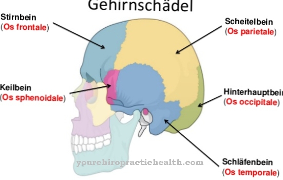
Under the Ethmoid bone the doctor understands a multi-limbed cranial skull bone of the bony eye socket. The ethmoid bone is involved in the anatomical structure of the eye sockets as well as the nasal and frontal sinuses and serves as a starting point for the olfactory system. The ethmoid bone can be affected by fractures, inflammation, and nerve damage.
What is the ethmoid bone
The ethmoid is a small, light and outwardly invisible cranial skull bone. This anatomical structure is also known as the ethmoid bone and lies at the end of the nasal cavity. There it forms the border to the cranial cavity in the depths. Thus, the ethmoid is a part of the base of the skull, but also of the roof of the nose and the orbit.
The bone consists of several sections: the lamina cribrosa, the lamina perpendicularis and the paired labyrinth. Each of these sections has a different function. The ethmoid bone is often referred to as a perforated bone plate, through whose holes the nerve cords of the olfactory brain run towards the nose. In this context, the separation of the nasal cavity and the cranial cavity is often mentioned as the main task of the anatomical structure.
Anatomy & structure
The lamina cribrosa is one of the four parts of the ethmoid bone. A two-winged bone protrusion protrudes from its center. This ledge is also known as the Hahnenkamm. One of the edges of this cockscomb articulates with the frontal bone. Its two wings correspond to the indentations of the frontal bone and thus form the blind opening in the tissue of the frontal sinus.
The perpendicular plate is the second structure of the ethmoid bone. This bone lamella forms the nasal septum. The second bone of the ethmoid also articulates with the nasal bone, the sphenoid bone, and the ploughshare bone. The two-part and symmetrically laid out labyrinth is the third structure of the ethmoid bone that carries different types of so-called ethmoid cells.
The labyrinth is involved as a structure both on the walls of the orbeitae and the nasal wall, as well as on the sphenoid bone. Overall, the surface of the ethmoid bone is rather smooth. Only the starting points of the individual nerves and blood vessels are not smooth in structure.
Function & tasks
The ethmoid bone is primarily responsible for the stability of the bony eye socket. It serves as a connecting piece between your individual structures of the olfactory bulb, eye socket and forehead area. The structural separation is also the task of the ethmoid bone. For example, the bone structures of the ethmoid separate the cranial cavity from the nasal cavity.
One edge of the cockscomb is also the starting point for the separating structure of the two cerebral hemispheres. Likewise, the two sides of the nose are divided by the ethmoid bone. This plays an important role in smell perception. It is only through the two nasal cavities that a person can, for example, assess in which direction the source of an odor is located. It is not only because of this function that the ethmoid bone plays an important role for the entire olfactory system and general olfactory perception. The second ethmoid bone serves as a starting point for many olfactory nerves in the upper area.
Without the holes in the ethmoid plate, the olfactory nerve and the blood vessels of the mucous membrane could not even penetrate into the nose. On the sides of the cockscomb, the first ethmoid bone also has a pit to support the right and left olfactory bulbs and through the fine tubules in this structure the olfactory nerve fibers extend into the olfactory bulb.
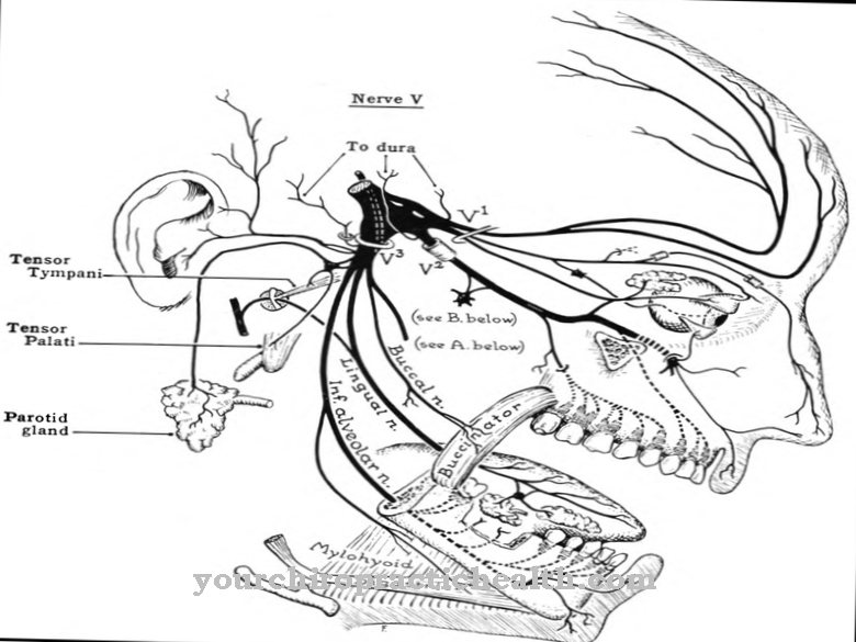
The nasociliary nerve, that is, part of the fifth cranial nerve, also runs through a notch in the first ethmoid bone. The fifth cranial nerve is responsible, among other things, for the transmission of stimuli between the eyes, upper jaw, lower jaw and brain and thus enables, for example, the chewing movement when eating.
Diseases
One of the most common ailments of the ethmoid bone is a fracture. If there is a fracture in one of the structures involved, it is usually associated with a blow to the eye socket. As a result, the ethmoid bone may run the risk of sagging. When this danger occurs, the bony eye socket and the nasal wall may no longer be stable.
A fracture of the ethmoid can possibly be corrected surgically or minimally invasively. If such a correction does not take place, the anatomical structure of the face can shift permanently from the frontal sinus downwards. Since the fifth cranial nerve and the olfactory nerves dock at the level of the ethmoid bone, nerves are sometimes involved in a fracture of the ethmoid bone. The nerves of the olfactory system are most commonly affected. This phenomenon can, for example, cause confused olfactory perceptions. Surgical liberation of the affected nerve structures is essential in this case. However, nerves that have been trapped for longer usually die.
This death causes permanent impairment of nerve function. Even the release of a pinched nerve may not be able to fully restore its functionality. In addition to fractures, the ethmoid bone and especially the ethmoid bone cells can also be affected by inflammatory processes. Such an inflammation is also known as "ethmoid sinusitis". Because of the connection to the cerebral hemispheres, inflammation of the ethmoid cells often spreads to the meninges and can thus cause meningitis. Just as often, if the course is unfavorable, inflammation of the ethmoid cells leads to an abscess of the orbit.


.jpg)
