The Occiput (Occipital bone) is part of the brain skull. The bone consists of three parts and not only contains various openings, but also serves tissue as a point of attachment. A skull base fracture can break the occiput and trisomy 18 often results in a large occiput.
What is the occiput
The bones of the skull capsule form a rounded vault that contains the brain. They provide support for the soft tissue of the complex organ and shield it from direct contact with the environment. The occiput is one of the bones that make up the brain skull (neurocranium). In total, the skull has seven different bones, the entire skull - including the facial skull - comprises 22.
The occiput is at the back of the head, where it can be found between the sphenoid bone (os sphenoidale), temple bone (os temporale) and parietal bone (os parietale). The anatomy also knows the occiput under the technical term "Os occipitale". Like all bones, the flat skull bone consists of a framework made of tissue that only hardens completely in the course of physical development.
Anatomy & structure
The occiput consists of three parts that are usually fused together: the pars squamosa, the pars lateralis and the pars basilaris. The pars squamosa lies below (dorsal) the foramen magnum.
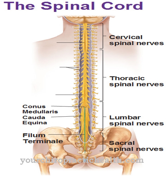
The foramen magnum is a large opening in the skull through which the elongated medulla (medulla oblongata) leaves the posterior cranial fossa (fossa cranii posterior) and merges into the spinal cord. The pars squamosa has a bowl-shaped shape and develops from two subunits. The occipital plate arises from four centers from where the bone tissue grows together. In contrast, the neck plate of the pars squamosa develops from two nuclei from the seventh week of development.
The pars lateralis forms the lateral areas of the occiput and arises from a nucleus about a week later. On each side the pars lateralis has a condylus occipitalis, which is part of the atlanto-occipital joint (articulatio atlantooccipitalis). The pars basilaris forms the part of the occiput that closes the skull rostrally towards the middle of the head. It has approximately the shape of a square and also arises from a center during physical development.
Function & tasks
As part of the skull, the occiput has the task of supporting and shielding the brain. It also contains or supports numerous structures.
Together with the temporal bone, the occiput forms the posterior fossa. It contains the cerebellum, the midbrain, the bridge and the elongated marrow. The latter protrudes through the foramen magnum, which is located at the bottom of the occiput. The pars squamosa has protrusions and depressions in the bones. One such depression is the transverse sinus sulcus, in which the transverse sinus runs. The transverse sinus is a blood conductor that draws venous blood from the skull.
Another indentation in the pars squamosa of the occiput is the sulcus sinus sigmoidei. It contains the sigmoid sinus, another venous blood conductor. The two sulci are located on the inside of the pars squamosa. There the Protuberantia occipitalis interna forms a small protrusion on which there is an attachment of the cerebral sickle (Falx cerebri). The skin separates the two halves of the brain. On the outside of the pars squamosa, the protuberantia occipitalis externa also offers a starting point for the hood or trapezius muscle (trapezius muscle).
At the pars lateralis of the occiput, the skull is connected to the atlas via the atlanto-occipital joint. The atlas represents the uppermost cervical vertebra (C1) and thus forms the beginning of the spine. On the inside of the pars lateralis is the jugular tubercle, which covers the hypoglossal canal as a protruding bone. In some cases the jugular tubercle also provides a recess for the cranial nerves IX-XI. With the help of its jugular process, the pars lateralis also functions as the starting point of a neck muscle, the muscle rectus capitis lateralis.
Furthermore, the occiput forms an inner protrusion on the pars lateralis, which is known as the pharyngeal tubercle. This is where the rectus capitis anterior muscle, the pharyngeal suture (raphe pharyngis) and the longus capitis muscle begin. The clivus of the pars lateralis forms the border between the fossa cranii posterior and the fossa cranii media.
Diseases
Injuries to the head can lead to a fracture of the base of the skull, which often also affects the occiput. Medicine distinguishes between a frontobasal fracture with involvement of the nose and a laterobasal fracture in which the temporal bone also breaks.
Possible symptoms include monocular / eyeglass hematoma, leakage of cerebrospinal fluid and blood, and impaired consciousness. If cranial nerves or parts of the brain are damaged, additional neurological symptoms may appear, such as the failure of cranial nerves. However, some symptoms can also resemble the clinical picture of a stroke. In some cases, the fracture of the base of the skull causes bleeding in the area of the eye. Affected people may feel a pulsation in the eye or the swelling of the eyeball pushes forward. Doctors then speak of an exophthalmos or protrusio bulbi.
In connection with trisomy 18, the occiput of those affected is often noticeably large. The genetic disorder is also known as Edwards syndrome and it can manifest itself in very different ways. Malformations and short stature are particularly typical. In most cases (around 90%), trisomy 18 leads to death before birth, and the mortality rate of children born with Edwards syndrome is also very high. Treatment usually focuses on the symptoms as medicine cannot treat the cause of the genetic disease. In some cases, supportive measures are necessary, for example artificial nutrition.




.jpg)

.jpg)
.jpg)


.jpg)