Tight junctions are protein networks. They girdle the endothelial tissue of the intestine, bladder and brain and, in addition to stabilizing functions, also take on barrier functions. Disturbances of these barrier functions have a negative effect on the different milieus of the body.
What is a tight junction?
Each cell membrane contains different proteins. The individual membrane proteins form a more or less dense network. In this context, a "tight junction", called "Zonula occludens" in Latin and "tight junction" in English, is a kind of protein-containing terminal strip that girdles the epithelial cells of vertebrates, for example, and is closely connected to neighboring cell ligaments.
Tight junctions seal the spaces between cells. They correspond to a barrier to diffusion. Diffusion is a substance transport path in the body of living beings that absorbs individual molecules into the cells. In the form of a diffusion barrier, tight junctions control the flow of molecules into the epithelium. In addition, they prevent the diffusion of membrane components from the apical into the lateral area and vice versa. Through the latter function, they maintain the polarity of the epithelial cells.
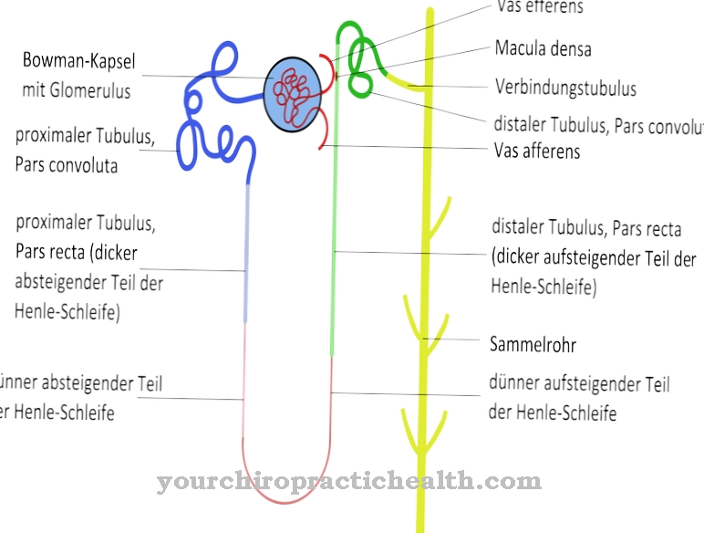
Tight junctions girdle the kidney, urinary bladder and intestinal epithelium. In addition, they are a functional component of the so-called blood-brain barrier and ensure that substances from the blood cannot diffuse into the tissue of the brain. The final ridges made of membrane proteins can contain various proteins. Probably not all of them are known yet.
Anatomy & structure
The most important membrane proteins within tight junctions are claudins and occludin. Claudins have been documented in more than 20 different vertebrates. All integral membrane proteins have a net-like arrangement and connect the membranes of several cells in a head-to-head contact. Watery pores make up the anatomy.
The composition of the membrane proteins contained differs from epithelium to epithelium and depends on the functional requirements of the tight junctions. For example, the claudin 16 in the renal epithelium is involved in the uptake of Mg2 + ions by the kidneys into the blood. Tight junctions form networks of different tightness depending on the task and the epithelium. The membrane proteins sit loosely in the intestine. The blood-brain barrier forms a relatively tight barrier.
The tightness of the network correlates with the permeability. The protein network consists of narrow strands. Especially the extracellular areas of the individual proteins combine to form a cell connection. The intracellular areas are attached to the cytoskeleton of cells. The tight junctions surround the cell circumference of an epithelium like a belt and thus nestle against the epithelial cell association.
Function & tasks
Tight junctions are primarily a diffusion barrier. This function can hold back molecules entirely from the intracellular space or be associated with a selective permeability (semipermeability) for molecules of a certain size. The network of tight junctions, through its function as a diffusion barrier, is the prerequisite for transcytosis. The paracellular diffusion of molecules or ions through the epithelial space is prevented by the tight junctions.At the same time, the end strips keep body fluids from flowing out.
The membrane proteins of the tight junctions also protect the organism from invading microorganisms and thus also form a barrier for living intruders. In addition to the barrier function, tight junctions also have a so-called fence function. The protein network prevents the movement of individual membrane components and thus maintains the cell polarity of the epithelium. The epithelium is divided into apical and basal areas by the networks. The apical cell membrane of the epithelium has a different biochemistry than the basolateral cell membrane. The tight junctions help to maintain these biochemical milieu differences and thereby enable a directed transport of substances.
Mechanical functions are added to these functions. For example, tight junctions also serve to stabilize epithelial cell assemblies. They connect the cells of the cytoskeleton with each other and ensure the tissue statics of the epithelium. The permeability between the epithelial cells is subject to temporary changes. The epithelium is thus able to react to increased paracellular transport requirements. To this end, the claudins and occludins of the "tight junctions" associate with the intracellular membrane proteins that connect to the actin cytoskeleton.
You can find your medication here
➔ Medicines for muscle weaknessDiseases
The tight junctions can be subject to changes in structure due to mutations and thus lose their functions. The claudin 16 of the protein networks in the kidney epithelium is not present in the required form after mutations in the protein-coding gene. Such mutations can result in a loss of Mg2 +.
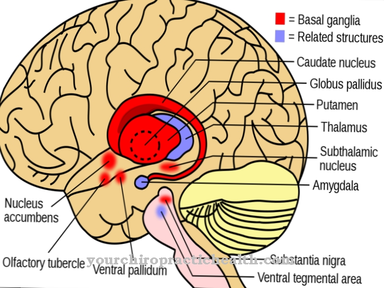
Because of the loss of the barrier function, too few Mg2 + ions are absorbed from the kidneys into the blood and too many are excreted with the urine. Diseases can also affect the "zonula occludens". This is especially true of the brain. The blood-brain barrier is a natural diffusion barrier between the blood and the brain that maintains the brain's milieu. Disorders of the blood-brain barrier occur, for example, in the context of multiple sclerosis. However, diseases such as diabetes mellitus can also disrupt the blood-brain barrier. The protective effect of the barrier is also lost with various brain injuries and degenerative diseases.
In multiple sclerosis, it is the recurring inflammation of the brain that has a harmful effect on the tight junctions. The cells of the body's own immune defense overcome the blood-brain barrier as part of the autoimmune disease. In an ischemic stroke, components of the tight junctions within the blood-brain barrier are even broken down. This type of stroke is accompanied by an emptiness in the brain, which is then refilled with blood. The endothelia of the blood-brain barrier change in two phases.
Since oxidants, proteolytic enzymes and cytokines are released by the pathological process, the permeability of the blood-brain barrier changes. Edema develops in the brain. Activated leukocytes then release so-called matrix metalloproteases, which lead to the breakdown of the basal lamina and protein complexes in the tight junctions.

